Beckham SA,
Matak MY,
Belousoff MJ,
Venugopal H,
Shah N,
Vankadari N,
Elmlund H,
Nguyen JHC,
Semler BL,
Wilce MCJ
Wilce JA,
Nucleic Acids Res
(2020)
Europe PMC
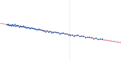
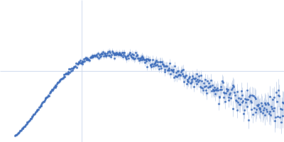
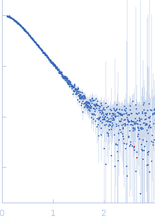
Synchrotron SAXS data from solutions of PCBP2 in 5 mM HEPES-KOH, 25 mM KCl, 2 mM MgCl2, 2 mM DTT, 4 % glycerol, 0.1 mM EDTA, pH 7.5 were collected on the SAXS/WAXS beam line at the Australian Synchrotron (Melbourne, Australia) using a Pilatus 1M detector at a sample-detector distance of 2.7 m and at a wavelength of λ = 0.10322 nm (I(s) vs s, where s = 4πsinθ/λ, and 2θ is the scattering angle). In-line size-exclusion chromatography (SEC) SAS was employed. The SEC parameters were as follows: A 100 μl sample at 0.5 mg/ml was injected at a 0.40 ml/min flow rate onto a GE Superdex 200 Increase 10/300 column at 15°C. 720 successive 5 second frames were collected through the SEC elution. The data were normalized to the intensity of the transmitted beam and radially averaged; the scattering of the solvent-blank was subtracted.
 s, nm-1