Beckham SA,
Matak MY,
Belousoff MJ,
Venugopal H,
Shah N,
Vankadari N,
Elmlund H,
Nguyen JHC,
Semler BL,
Wilce MCJ,
Wilce JA,
Nucleic Acids Res
(2020)
Europe PMC
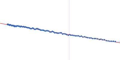
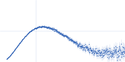
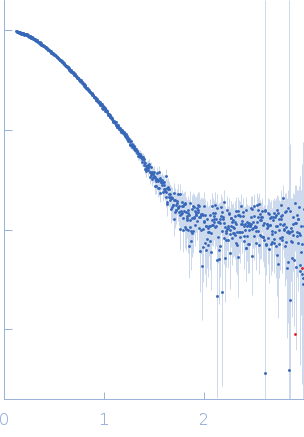
SASDH95 – Truncated Poly(rC)-binding protein 2 (PCBP2-ΔKH3)
Synchrotron SAXS data from solutions of Poly(rC)-binding protein 2 (PCBP2) ΔKH3 in 5 mM HEPES-KOH, 25 mM KCl, 2 mM MgCl2, 2 mM DTT, 4 % glycerol, 0.1 mM EDTA, pH 7.5 were collected on the SAXS/WAXS beam line at the Australian Synchrotron (Melbourne, Australia) using a Pilatus 1M detector at a sample-detector distance of 2.7 m and at a wavelength of λ = 0.10322 nm (I(s) vs s, where s = 4πsinθ/λ, and 2θ is the scattering angle). In-line size-exclusion chromatography (SEC) SAS was employed. The SEC parameters were as follows: A 100 μl sample was injected at a 0.40 ml/min flow rate onto a GE Superdex 200 Increase 10/300 column at 15°C. 720 successive 5 second frames were collected through the SEC elution. The data were normalized to the intensity of the transmitted beam and radially averaged; the scattering of the solvent-blank was subtracted.
 s, nm-1