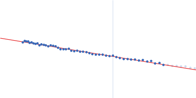
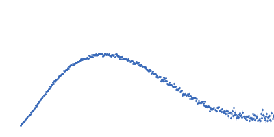
Dmax unknown – experimental data range validation not possible.
There are no models related to this curve.
Synchrotron SAXS
data from solutions of
N-terminal engineered, disulfide-containing endo-beta-N-acetylglucosaminidase H at 1161.6 Gy X-ray dose (J/kg)
in
20 mM Tris-HCl, 50 mM NaCl, 5 mM EDTA, pH 7.5
were collected
on the
12.3.1 (SIBYLS) beam line
at the Advanced Light Source (ALS) storage ring
(Berkeley, CA, USA)
using a Pilatus3 X 2M detector
at a sample-detector distance of 2 m and
at a wavelength of λ = 0.1127 nm
(I(s) vs s, where s = 4πsinθ/λ, and 2θ is the scattering angle).
One solute concentration of 5.00 mg/ml was measured
at 10°C.
One
0.300 second frame was collected.
The data were normalized to the intensity of the transmitted beam and radially averaged; the scattering of the solvent-blank was subtracted.
Molecular weight was estimated from the volume of correlation (Vc; Rambo & Tainer 2013). Dose was estimated using RADDOSE modified for SAXS (Brooks-Barlett et al., 2017).
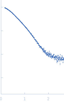 s, nm-1