Alternative dimerization is required for activity and inhibition of the HEPN ribonuclease RnlA

Garcia-Rodriguez G, Charlier D, Wilmaerts D, Michiels J, Loris R, Nucleic Acids Research 49(12):7164-7178 (2021) DOI

SASDHX7 – Escherichia coli RnlA (mRNA endoribonuclease toxin LS)

mRNA endoribonuclease toxin LS
MWexperimental 85 kDa
MWexpected 80 kDa
VPorod 134 nm3
log I(s) 4.99×101 4.99×100 4.99×10⁻¹ 4.99×10⁻²
mRNA endoribonuclease toxin LS small angle scattering data  s, nm-1
ln I(s)
mRNA endoribonuclease toxin LS Guinier plot ln 5.00×101 Rg: 3.7 nm 0 (3.7 nm)-2 s2
(sRg)2I(s)/I(0)
mRNA endoribonuclease toxin LS Kratky plot 1.104 0 3 sRg
p(r)
mRNA endoribonuclease toxin LS pair distance distribution function Rg: 3.6 nm 0 Dmax: 10.7 nm

Data validation


There are no models related to this curve.

Synchrotron SAXS data from solutions of Escherichia coli RnlA (mRNA endoribonuclease toxin LS) in 20 mM Tris, 150 mM NaCl, 1 mM TCEP, 5% glycerol, pH 8 were collected on the BM29 beam line at the ESRF storage ring (Grenoble, France) using a Pilatus 1M detector at a sample-detector distance of 2.8 m and at a wavelength of λ = 0.09919 nm 
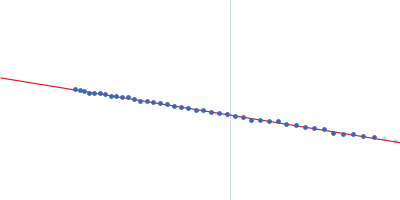
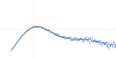
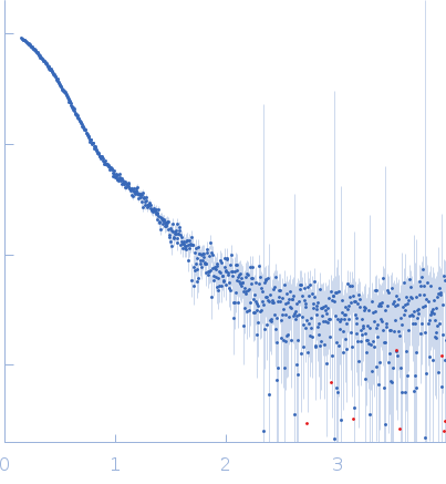
(I(s) vs s, where s = 4πsinθ/λ, and 2θ is the scattering angle). Solute concentrations ranging between 1.7 and 2.4 mg/ml were measured at 20°C. 10 successive 1 second frames were collected. The data were normalized to the intensity of the transmitted beam and radially averaged; the scattering of the solvent-blank was subtracted. The low angle data collected at lower concentrations were extrapolated to infinite dilution and merged with the higher concentration data to yield the final composite scattering curve.

mRNA endoribonuclease toxin LS (RnlA)
Mol. type   Protein
Organism   Escherichia coli
Olig. state   Dimer
Mon. MW   40.1 kDa
 
UniProt   P52129 (1-357)
Sequence   FASTA