Structural Investigations of Human A2M Identify a Hollow Native Conformation That Underlies Its Distinctive Protease-Trapping Mechanism.

Harwood SL, Lyngsø J, Zarantonello A, Kjøge K, Nielsen PK, Andersen GR, Pedersen JS, Enghild JJ, Mol Cell Proteomics 20:100090 (2021) Europe PMC

SASDJL3 – Methylamine-treated alpha-2-macroglobulin, glycosylated

Alpha-2-macroglobulin
MWI(0) 681 kDa
MWexpected 643 kDa
log I(s) 7.80×10⁻¹ 7.80×10⁻² 7.80×10⁻³ 7.80×10⁻⁴
Alpha-2-macroglobulin small angle scattering data  s, nm-1
ln I(s)
Alpha-2-macroglobulin Guinier plot ln 7.80×10⁻¹ Rg: 6.7 nm 0 (6.7 nm)-2 s2
(sRg)2I(s)/I(0)
Alpha-2-macroglobulin Kratky plot 1.104 0 3 sRg
p(r)
Alpha-2-macroglobulin pair distance distribution function Rg: 6.5 nm 0 Dmax: 19.7 nm

Data validation


Fits and models


log I(s)
 s, nm-1
Alpha-2-macroglobulin CUSTOM IN-HOUSE model

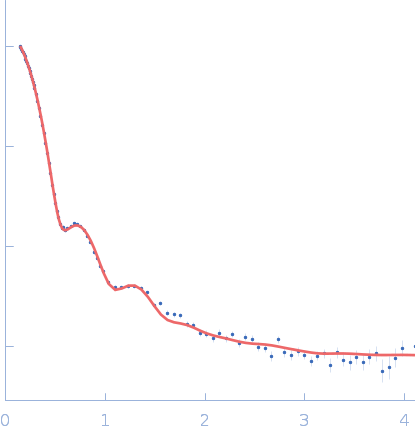
SAXS data from a solution of human methylamine-treated glycosylated alpha-2-macroglobulin (molecular mass of roughly 720 kDa for the tetramer of 180 kDa subunits) in a 20 mM HEPES, 150 mM NaCl, pH 7.4, buffer were collected at the in-house Bruker AXS NanoSTAR instrument with an Excillum Ga Metal Jet X-ray source at Aarhus University (I(s) vs s, where s = 4π sin θ/λ; 2θ is the scattering angle; λ = 0.134 nm). The instrument has a homebuilt scatterless pinhole in front of the sample and is equipped with a Bruker AXS Vantec2000 detector. Data were collected for 1800 s for the sample and buffer, respectively, and the data were normalized to absolute scale using the scattering from pure water. The structure was modelled using home-written software, which performs rigid body refinement on the structure with P222 symmetry and takes into account a hydration layer.
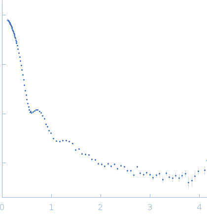
Alpha-2-macroglobulin (A2M)
Mol. type   Protein
Organism   Homo sapiens
Olig. state   Tetramer
Mon. MW   160.8 kDa
 
UniProt   P01023 (24-1474)
Sequence   FASTA