Dmax unknown – experimental data range validation not possible.
There are no models related to this curve.
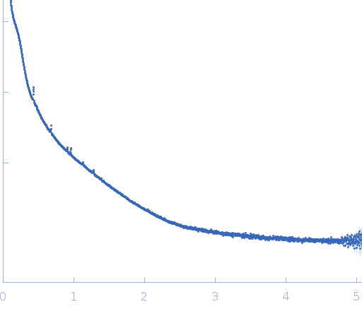
Synchrotron SAXS data from Sf9 cell cultures containing in cellulo IMPDH protein crystals were collected on the EMBL P12 beam line at PETRA III (DESY; Hamburg, Germany) using a Pilatus 2M detector at a sample-detector distance of 3 m and at a wavelength of λ = 0.124 nm (I(s) vs s, where s = 4πsinθ/λ, and 2θ is the scattering angle). Data were collected at 20°C using a fixed sample position (no sample flow) in a 1.8 mm capillary. 40 successive 0.045 second frames were collected; the individual 2D images were summed (1.8 s total exposure) and then normalized to the intensity of the transmitted beam and radially averaged. The scattering of the solvent-blank was subtracted.
The Sf9 cell cultures were suspended in TBS (20 mM Tris, 150 mM NaCl), pH 7, that was used for solvent-background scattering subtraction. Data validation metrics do not apply for this entry (Rg, I(0), MW, etc). The quoted 'experimental MW' is that of the monomeric protein calculated from the amino acid sequence.
 s, nm-1