Crystal and solution structures of a novel antimicrobial peptide from Chrysomya megacephala.

Xiao C, Xiao Z, Hu C, Lu J, Cui L, Zhang Y, Dai Y, Zhang Q, Wang S, Liu W, Acta Crystallogr D Struct Biol 77(Pt 7):894-903 (2021) Europe PMC

SASDKX6 – PSK, an antimicrobial peptide from Chrysomya megacephala

PSK, an antimicrobial peptide from Chrysomya megacephala
MWexperimental 11 kDa
MWexpected 10 kDa
VPorod 18 nm3
log I(s) 6.06×100 6.06×10⁻¹ 6.06×10⁻² 6.06×10⁻³
PSK, an antimicrobial peptide from Chrysomya megacephala small angle scattering data  s, nm-1
ln I(s)
PSK, an antimicrobial peptide from Chrysomya megacephala Guinier plot ln 6.06×100 Rg: 1.6 nm 0 (1.6 nm)-2 s2
(sRg)2I(s)/I(0)
PSK, an antimicrobial peptide from Chrysomya megacephala Kratky plot 1.104 0 3 sRg
p(r)
PSK, an antimicrobial peptide from Chrysomya megacephala pair distance distribution function Rg: 1.7 nm 0 Dmax: 5 nm

Data validation


Fits and models


log I(s)
 s, nm-1
PSK, an antimicrobial peptide from Chrysomya megacephala DAMMIF model

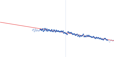
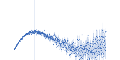
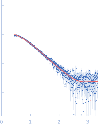
Synchrotron SAXS data from solutions of the antimicrobial PSK-peptide from Chrysomya megacephala, in 20 mM Tris, 150 mM NaCl, 1 mM DTT, pH 8 were collected on the BL19U2 beam line at the Shanghai Synchrotron Radiation Facility (SSRF, Shanghai, China) using a Pilatus 1M detector at a wavelength of λ = 0.103 nm (I(s) vs s, where s = 4πsinθ/λ, and 2θ is the scattering angle). One solute concentration of 1.00 mg/ml was measured at 10°C. 23 successive 1 second frames were collected. The data were normalized to the intensity of the transmitted beam and radially averaged; the scattering of the solvent-blank was subtracted.

Sample detector distance = UNKNOWN
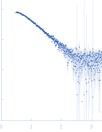
PSK, an antimicrobial peptide from Chrysomya megacephala (PSK)
Mol. type   Protein
Organism   Chrysomya megacephala
Olig. state   Monomer
Mon. MW   10.4 kDa
Sequence   FASTA