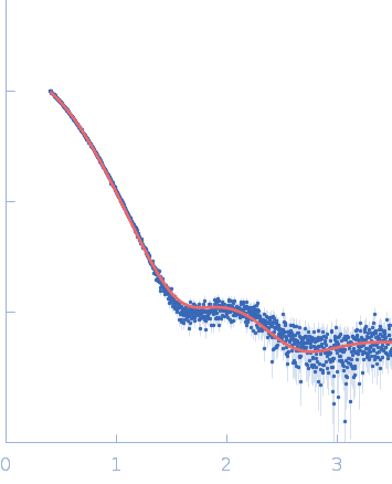
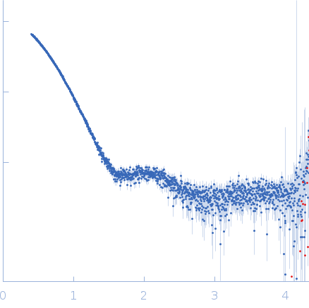
Dual Role of the Active Site Residues of Thermus thermophilus 3-Isopropylmalate Dehydrogenase: Chemical Catalysis and Domain Closure.

Gráczer É, Szimler T, Garamszegi A, Konarev PV, Lábas A, Oláh J, Palló A, Svergun DI, Merli A, Závodszky P, Weiss MS, Vas M, Biochemistry 55(3):560-74 (2016) Europe PMC

SASDL67 – Mutation of the Active Site Residues of Thermus thermophilus 3‑Isopropylmalate Dehydrogenase

3-isopropylmalate dehydrogenase
MWI(0) 72 kDa
MWexpected 73 kDa
log I(s) 1.46×10⁴ 1.46×10³ 1.46×10² 1.46×101
3-isopropylmalate dehydrogenase small angle scattering data  s, nm-1
ln I(s)
3-isopropylmalate dehydrogenase Guinier plot ln 1.46×10⁴ Rg: 2.8 nm 0 (2.8 nm)-2 s2
(sRg)2I(s)/I(0)
3-isopropylmalate dehydrogenase Kratky plot 1.104 0 3 sRg

Data validation


Fits and models


log I(s)
 s, nm-1
3-isopropylmalate dehydrogenase PDB (PROTEIN DATA BANK) model

Synchrotron SAXS data from solutions of Mutation of the Active Site Residues of Thermus thermophilus 3‑Isopropylmalate Dehydrogenase in 25 mM MOPS/NaOH, pH 7.6 were collected on the EMBL P12 beam line at the PETRA III storage ring (DESY; Hamburg, Germany) using a Pilatus 2M detector at a sample-detector distance of 3.1 m and at a wavelength of λ = 0.124 nm (I(s) vs s, where s = 4πsinθ/λ, and 2θ is the scattering angle). One solute concentration of 4.52 mg/ml was measured at 10°C. 20 successive 0.050 second frames were collected. The data were normalized to the intensity of the transmitted beam and radially averaged; the scattering of the solvent-blank was subtracted.

The OLIGOMER analysis is made available in the full entry zip archive comparing for the D217A, D245A, K185A, N102A, and Y139A mutant state(s) in solution.

3-isopropylmalate dehydrogenase (Tt-IPMDH)
Mol. type   Protein
Organism   Thermus thermophilus
Olig. state   Dimer
Mon. MW   36.7 kDa
 
UniProt   P61495 (1-345)
Sequence   FASTA
 
PDB ID   2Y3Z
 
PDB ID   4F7I