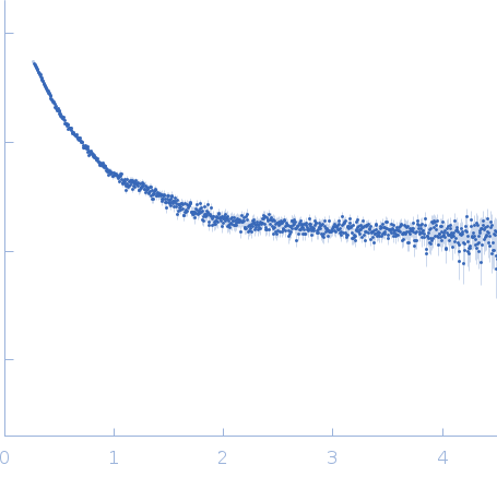
Conformational landscape of multidomain SMAD proteins

Gomes T, Martin-Malpartida P, Ruiz L, Aragón E, Cordeiro T, Macias M, Computational and Structural Biotechnology Journal (2021) DOI

SASDLA2 – Mothers against decapentaplegic homolog 2, SMAD2 phosphomimetic mutant 0.5 mg/ml

Mothers against decapentaplegic homolog 2 (C-terminus phosphomimetic mutant)
MWexperimental 97 kDa
MWexpected 53 kDa
log I(s) 1.37×10² 1.37×101 1.37×100 1.37×10⁻¹
Mothers against decapentaplegic homolog 2 (C-terminus phosphomimetic mutant) small angle scattering data  s, nm-1
ln I(s)
Mothers against decapentaplegic homolog 2 (C-terminus phosphomimetic mutant) Guinier plot ln 1.38×10² Rg: 5.1 nm 0 (5.1 nm)-2 s2
(sRg)2I(s)/I(0)
Mothers against decapentaplegic homolog 2 (C-terminus phosphomimetic mutant) Kratky plot 1.104 0 3 sRg
Dmax: 20 nm

Data validation


There are no models related to this curve.

Synchrotron SAXS data from solutions of Mothers against decapentaplegic homolog 2, SMAD2 phosphomimetic mutant 0.5 mg/ml in 20 mM Tris, 150 mM NaCl, pH 7.2 were collected on the BM29 beam line at the ESRF storage ring (Grenoble, France) using a Pilatus 1M detector (I(s) vs s, where s = 4πsinθ/λ, and 2θ is the scattering angle). One solute concentration of 0.50 mg/ml was measured at 10°C. The data were normalized to the intensity of the transmitted beam and radially averaged; the scattering of the solvent-blank was subtracted.

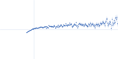
Monomer-dimer trimer equilibrium. CAUTION! Severe low-s truncation; Guinier region is not present! X-ray wavelength: UNKNOWN. Sample-to-detector distance: UNKNOWN. X-ray exposure time: UNKNOWN. PED: https://proteinensemble.org/PED00200

Tags: idp PED
Mothers against decapentaplegic homolog 2 (C-terminus phosphomimetic mutant)
Mol. type   Protein
Organism   Homo sapiens
Olig. state   Other
Mon. MW   53.4 kDa
 
UniProt   Q15796 (1-463)
Sequence   FASTA