Integrative modeling of guanylate binding protein dimers

Schumann W, Loschwitz J, Reiners J, Degrandi D, Legewie L, Stühler K, Pfeffer K, Poschmann G, Smits S, Strodel B, Protein Science (2023) DOI

SASDLY9 – Mouse guanylate-binding protein 7 - mGBP7

Gbp6 protein (Gbp7 - Gbp6 protein Mus musculus)
MWI(0) 137 kDa
MWexpected 148 kDa
VPorod 176 nm3
log I(s) 9.92×10⁻³ 9.92×10⁻⁴ 9.92×10⁻⁵ 9.92×10⁻⁶
Gbp6 protein (Gbp7 - Gbp6 protein Mus musculus) small angle scattering data  s, nm-1
ln I(s)
Gbp6 protein (Gbp7 - Gbp6 protein Mus musculus) Guinier plot ln 9.93×10⁻³ Rg: 5.4 nm 0 (5.4 nm)-2 s2
(sRg)2I(s)/I(0)
Gbp6 protein (Gbp7 - Gbp6 protein Mus musculus) Kratky plot 1.104 0 3 sRg
p(r)
Gbp6 protein (Gbp7 - Gbp6 protein Mus musculus) pair distance distribution function Rg: 5.1 nm 0 Dmax: 17 nm

Data validation


Fits and models


log I(s)
 s, nm-1
Gbp6 protein (Gbp7 - Gbp6 protein Mus musculus) GASBOR model

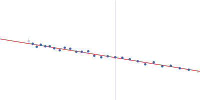
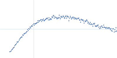
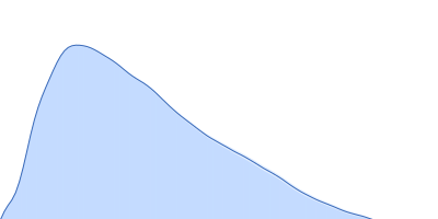
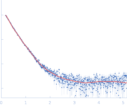
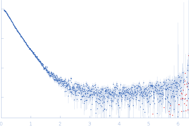
SAXS data from solutions of mouse guanylate-binding protein 7 (mGBP7) in 50 mM Tris, 5 mM MgCl, 2 mM DTT, pH 8 were collected using a Xenocs Xeuss 2.0 Q-Xoom instrument (Center for Structural Studies, Heinrich-Heine-University, Düsseldorf Germany) equipped with a Pilatus3 R 300K detector at a sample-detector distance of 0.6 m and at a wavelength of λ = 0.154 nm (I(s) vs s, where s = 4πsinθ/λ, and 2θ is the scattering angle). Solute concentrations ranging between 1.7 and 6.9 mg/ml were measured at 10°C. Six successive 600 second frames were collected. The data were normalized to the intensity of the transmitted beam and radially averaged; the scattering of the solvent-blank was subtracted. The low angle data collected at lower concentration were merged with the highest concentration high angle data to yield the final composite scattering curve.

Gbp6 protein (Gbp7 - Gbp6 protein Mus musculus) (mGbp7)
Mol. type   Protein
Organism   Mus musculus
Olig. state   Dimer
Mon. MW   73.8 kDa
 
UniProt   Q91Z40 (2-638)
Sequence   FASTA