Structural basis for the inhibition of the Bacillus subtilis c-di-AMP cyclase CdaA by the phosphoglucomutase GlmM

Pathania M, Tosi T, Millership C, Hoshiga F, Morgan R, Freemont P, Gründling A, Journal of Biological Chemistry :101317 (2021) DOI

SASDLZ4 – Bacillus subtilis phosphoglucosmine mutase GlmM

Phosphoglucosamine mutase
MWexperimental 85 kDa
MWexpected 101 kDa
VPorod 140 nm3
log I(s) 2.55×10⁻¹ 2.55×10⁻² 2.55×10⁻³ 2.55×10⁻⁴
Phosphoglucosamine mutase small angle scattering data  s, nm-1
ln I(s)
Phosphoglucosamine mutase Guinier plot ln 2.56×10⁻¹ Rg: 3.7 nm 0 (3.7 nm)-2 s2
(sRg)2I(s)/I(0)
Phosphoglucosamine mutase Kratky plot 1.104 0 3 sRg
p(r)
Phosphoglucosamine mutase pair distance distribution function Rg: 3.8 nm 0 Dmax: 12.2 nm

Data validation


There are no models related to this curve.

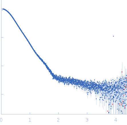
Synchrotron SAXS data from solutions of phosphoglucosmine mutase, GlmM, from Bacillus subtilis in 30 mM Tris, 150 mM NaCl, pH 7.5 were collected on the B21 beam line at the Diamond Light Source (Didcot, UK) using a Eiger 4M detector at a wavelength of λ = 0.1 nm (I(s) vs s, where s = 4πsinθ/λ, and 2θ is the scattering angle). In-line size-exclusion chromatography (SEC) SAS was employed. The SEC parameters were as follows: A 45.00 μl sample at 10 mg/ml was injected at a 0.16 ml/min flow rate onto a Shodex KW403 column at 15°C. 620 successive 3 second frames were collected through the entire elution profile. The data were normalized to the intensity of the transmitted beam and radially averaged; the scattering of the solvent-blank was subtracted from the SEC-peak sample scattering frames.

Sample detector distance = UNKNOWN

Phosphoglucosamine mutase (GlmM)
Mol. type   Protein
Organism   Bacillus subtilis (strain 168)
Olig. state   Dimer
Mon. MW   50.3 kDa
 
UniProt   O34824 (1-448)
Sequence   FASTA