|
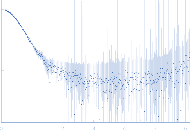
Synchrotron SAXS data from solutions of single-stranded DNA dC->dU-editing enzyme APOBEC3G in complex with a DNA inhibitor (dimer) in 50 mM phosphate pH 6.0, 200 mM NaCl, 2 mM β-mercaptoethanol (β-ME), 5% glycerol, 200 µM Na2-EDTA were collected on the SAXS/WAXS beam line at the Australian Synchrotron (Melbourne, Australia) using a Pilatus 1M detector at a sample-detector distance of 1.6 m and at a wavelength of λ = 0.10332 nm (I(s) vs s, where s = 4πsinθ/λ, and 2θ is the scattering angle). In-line size-exclusion chromatography (SEC) SAS was employed using a sheath-flow cell. The SEC parameters were as follows: A 100.00 μl sample at 1.3 mg/ml was injected at a 0.20 ml/min flow rate onto a GE Superdex 75 Increase 10/300 column at 25°C. 1000 successive 1 second frames were collected. The data were normalized to the intensity of the transmitted beam and radially averaged; the scattering of the solvent-blank was subtracted.
CAUTION! The bead model displayed in this entry is based on a two-phase bead modelling approach (e.g., macromolecule and solvent). However, the protein-DNA complex described for this entry consists of three phases (protein, DNA and solvent). Therefore the homogeneous scattering length density of the particle displayed here does not represent the shape/volume of the inhomogeneous scattering length density of the protein/DNA complex under investigation.
The displayed bead model (damfilt) shows the bead-occupancy and volume corrected spatial representation of the complex calculated from the alignment of several individual models. An individual model-fit is displayed to the left.
|
|
 s, nm-1