Structural basis of the zinc-induced cytoplasmic aggregation of the RNA-binding protein SFPQ

Huang J, Ringuet M, Whitten A, Caria S, Lim Y, Badhan R, Anggono V, Lee M, Nucleic Acids Research 48(6):3356-3365 (2020) DOI

SASDMG8 – Splicing factor, proline- and glutamine-rich-QM bound to Non-POU domain-containing octamer-binding protein: SFPQ(276-598)-QM / NONO(53-312) heterodimer

Non-POU domain-containing octamer-binding protein
Splicing factor, proline- and glutamine-rich
MWI(0) 62 kDa
MWexpected 68 kDa
VPorod 90 nm3
log I(s) 3.44×10⁻² 3.44×10⁻³ 3.44×10⁻⁴ 3.44×10⁻⁵
Non-POU domain-containing octamer-binding protein Splicing factor, proline- and glutamine-rich small angle scattering data  s, nm-1
ln I(s)
Non-POU domain-containing octamer-binding protein Splicing factor, proline- and glutamine-rich Guinier plot ln 3.45×10⁻² Rg: 4.0 nm 0 (4.0 nm)-2 s2
(sRg)2I(s)/I(0)
Non-POU domain-containing octamer-binding protein Splicing factor, proline- and glutamine-rich Kratky plot 1.104 0 3 sRg
p(r)
Non-POU domain-containing octamer-binding protein Splicing factor, proline- and glutamine-rich pair distance distribution function Rg: 4.2 nm 0 Dmax: 16.5 nm

Data validation


Fits and models


log I(s)
 s, nm-1
Non-POU domain-containing octamer-binding protein Splicing factor, proline- and glutamine-rich PDB (PROTEIN DATA BANK) model

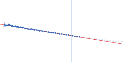
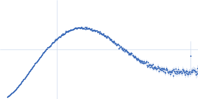
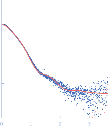
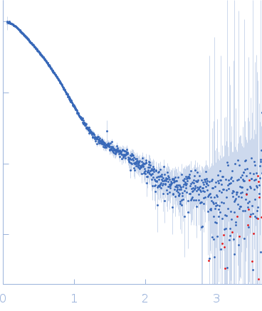
X-ray synchrotron radiation scattering data from solutions of human SFPQ(276-598)-QM/NONO(53-512) in 20 mM Tris 250 mM NaCl, pH 7.5 were collected on the SAXS/WAXS beam line of the Australian Synchrotron (Melbourne, Australia) using a 2D Photon counting Dectris/Pilatus 1M pixel detector (s = 4πsinθ/λ, where 2θ is the scattering angle). Here QM refers to the quadruple mutatation in the SFPQ(276-598) protein (L535A, L529A, L546A, M549A). Approximately 100 uL of sample at 0.75 mg/mL was drawn into a 1 mm capillary and as the sample passed through the beam, forty successive 1 second frames were collected. The data were normalized to the intensity of the transmitted beam, placed on an absolute scale relative to a water standard, and radially averaged. The scattering of the solvent-blank was subtracted to yield the scattering profile of the protein complex. The data was modelled using CRYSOL and PDB structure 4WIJ:B. Included in the attached zip archive are concentration series (0.38 - 3.0 mg/mL) for the protein in the absence and presence of Zn(II) (protein to zinc molar ratio of 1 : 0.5).

Non-POU domain-containing octamer-binding protein (NONO)
Mol. type   Protein
Organism   Homo sapiens
Olig. state   Monomer
Mon. MW   30.1 kDa
 
UniProt   Q15233 (53-312)
Sequence   FASTA
 
Splicing factor, proline- and glutamine-rich (SFPQ-QM)
Mol. type   Protein
Organism   Homo sapiens
Olig. state   Monomer
Mon. MW   38.0 kDa
 
UniProt   P23246 (276-598)
Sequence   FASTA
 
PDB ID   4WIJ