|
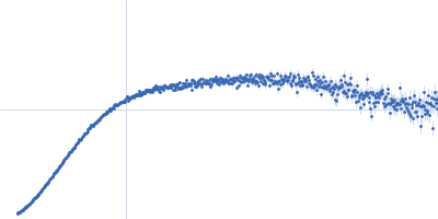
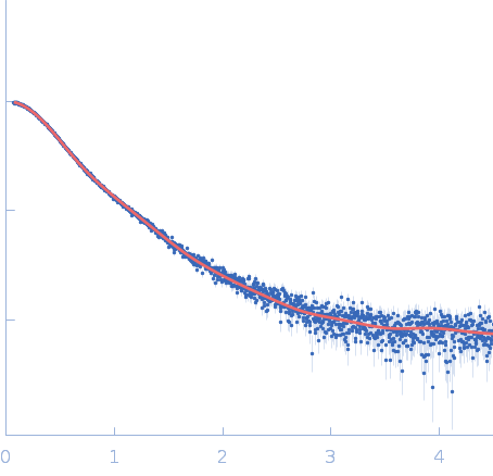
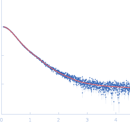
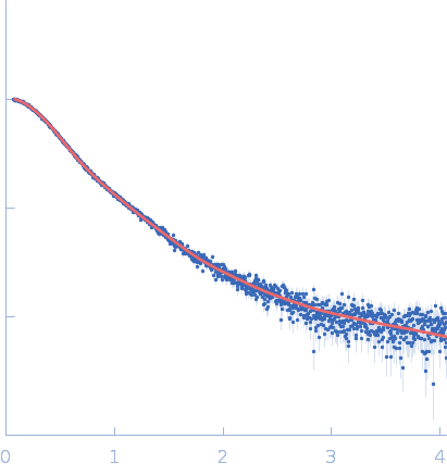
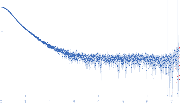
Synchrotron SAXS data from solutions of MITF in 50 mM HEPES, 150 mM NaCl, 10 mM CaCl2, pH 7.2 were collected on the EMBL P12 beam line at PETRA III (DESY, Hamburg, Germany) using a Pilatus 6M detector at a sample-detector distance of 3 m and at a wavelength of λ = 0.124 nm (I(s) vs s, where s = 4πsinθ/λ, and 2θ is the scattering angle). In-line size-exclusion chromatography (SEC) SAS was employed. The SEC parameters were as follows: A 50.00 μl sample at 5 mg/ml was injected at a 0.35 ml/min flow rate onto a GE Superdex 200 Increase 5/150 column at 20°C. 30 successive 1 second frames were collected through the sample elution peak. The data were normalized to the intensity of the transmitted beam and radially averaged; the scattering of the solvent-blank was subtracted using CHROMIXS software.
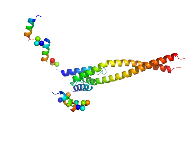
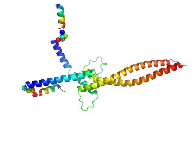
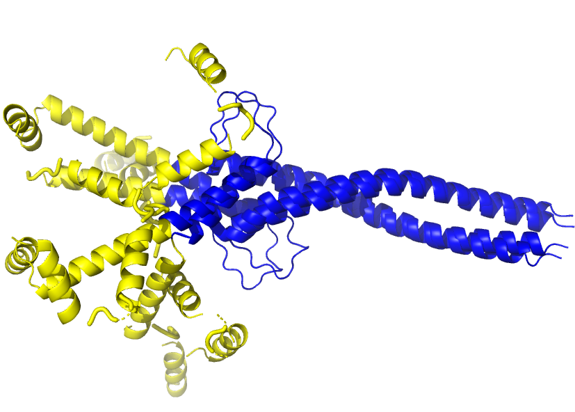
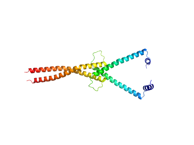
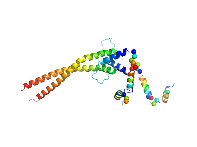
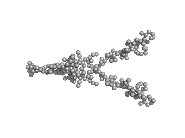
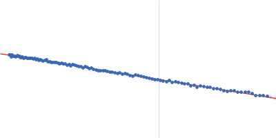
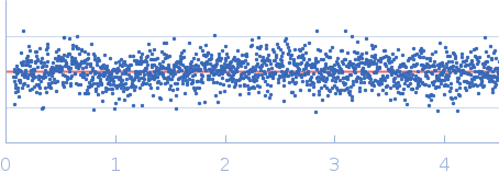
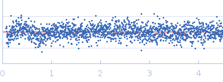
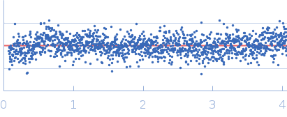
The SEC-SAXS data, the Rg correlation through the SEC-SAXS peak and alternative models of MITF are provided in the full entry zip archive. The models of MITF shown in this entry are: 1) Top - individual CORAL models and an example model fit to the data, with a representation of the MITF ensemble aligned to the coiled-coil domain (blue) showing the different spatial disposition of the N-terminal helical arms. 2) Middle - the MITF sample may also be described as a mixture of a hybrid Alpha-fold/X-ray crystal structure (AF-Q08874-F1; PDB: 4ATI, 4ATH) conformation (38% volume fraction) in combination with a model where the N-terminal arms have 'melted' (62% volume fraction). 3) Bottom - a GASBOR dummy residue model representation of MITF with enforced P2 symmetry.
|
|

 s, nm-1