|
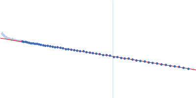
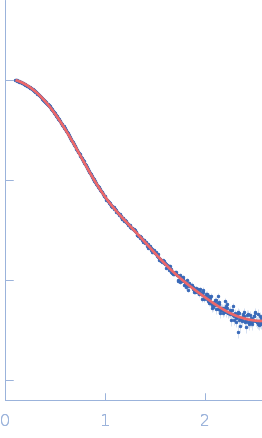
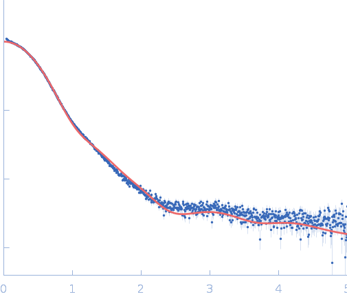
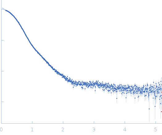
SAXS experiments for transferrin were carried out at BM29 beamline at the European Synchrotron Radiation Facility (ESRF, Grenoble, France). The wavelength of incident X-rays was 0.1 nm and the Pilatus3 2M detector was placed 2.87 m from the sample leading to a range of the scattering vector from 0.025 to 6 nm-1. To avoid radiation damage, samples with a volume of 30-40 µL were measured using a robotic sample handler in flow-through mode, collecting over 10 frames lasting 1 s for each sample. Frames were automatically checked for radiation damage and those not displaying any radiation damage were then averaged. Before and after each sample, buffer scattering was collected and subtracted from sample scattering. The buffer consisted of 15 mM HEPES, 20 mM NaHCO3, and 50 mM NaCl, tested at pH 8.0, 7.0, and 5.5. To assess concentration effects, a dilution series consisting of 2 concentrations (2.5 and 5 mg ml-1), for both Tf-apo and Tf-holo samples, was measured. Since the scattering curves for Tf did not display any concentration dependence at the highest concentrations, this concentration (5 mg ml-1) was used in our analysis. Merging, subtracting and subsequent analysis was performed using PRIMUS and Scatter software. A MX model (PDB ID: 2HAV) was compared with the SAXS data, including a modelling by GASBOR. For apo-Tf, the MX model fits reasonably well and non significant changes due the the pH were observed.
|
|
 s, nm-1