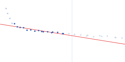
SANS data from cytochrome c' in phosphate buffer, pD 13, were collected using the KWS1 SANS instrument (FRM2, Munich, Germany) equipped with a 6Li-Scintillator 1 mm thickness + photomultiplier detector at a sample-detector distance of 4 m and at a wavelength of λ = 0.5 nm (I(s) vs s, where s = 4πsinθ/λ, and 2θ is the scattering angle). One solute concentration of 5.30 mg/ml was measured at 25°C. One 1800 second frame was collected. The data were normalized to the intensity of the transmitted beam and radially averaged; the scattering of the solvent-blank was subtracted. The low angle data collected at lower concentration were merged with the highest concentration high angle data to yield the final composite scattering curve.
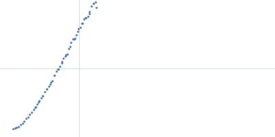
 s, nm-1
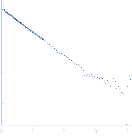