Inline small‐angle X‐ray scattering‐coupled chromatography under extreme hydrostatic pressure

Miller R, Cummings C, Huang Q, Ando N, Gillilan R, Protein Science 31(12) (2022) DOI

SASDP47 – Glucose Isomerase at a neutral pH and 5 MPa (1 atm) of pressure

Xylose isomerase
MWexperimental 170 kDa
MWexpected 173 kDa
VPorod 253 nm3
log I(s) 3.31×10⁻² 3.31×10⁻³ 3.31×10⁻⁴ 3.31×10⁻⁵
Xylose isomerase small angle scattering data  s, nm-1
ln I(s)
Xylose isomerase Guinier plot ln 3.32×10⁻² Rg: 3.4 nm 0 (3.4 nm)-2 s2
(sRg)2I(s)/I(0)
Xylose isomerase Kratky plot 1.104 0 3 sRg
Dmax: 10.6 nm

Data validation


There are no models related to this curve.

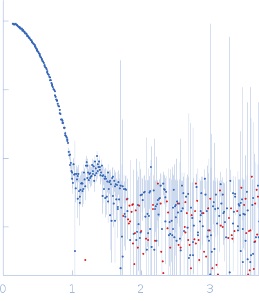
Synchrotron SAXS data from solutions of glucose isomerase in 25 mM HEPES, 150 mM NaCl, 3% v/v glycerol, pH 7 were collected on the ID7A1 BioSAXS / HP-Bio Beamline beam line at the Cornell High Energy Synchrotron Source (CHESS; Ithaca, NY, USA) using a Eiger 4M detector at a sample-detector distance of 1.6 m and at a wavelength of λ = 0.089 nm (I(s) vs s, where s = 4πsinθ/λ, and 2θ is the scattering angle). In-line size-exclusion chromatography (SEC) SAS was employed. The SEC parameters were as follows: A sample at 17.9 mg/ml was injected onto a column at 23°C. One 2 second frame was collected. The data were normalized to the intensity of the transmitted beam and radially averaged; the scattering of the solvent-blank was subtracted.

SEC column = UNKNOWN. Sample injection volume = UNKNOWN. Flow rate = UNKNOWN

Xylose isomerase
Mol. type   Protein
Organism   Streptomyces rubiginosus
Olig. state   Tetramer
Mon. MW   43.2 kDa
 
UniProt   P24300 (1-380)
Sequence   FASTA