Methionine gamma lyase fused with S3 domain VGF forms octamers and adheres to tumor cells via binding EGFR

Bondarev N, Bagaeva D, Bazhenov S, Buben M, Bulushova N, Ryzhykau Y, Okhrimenko I, Zagryadskaya Y, Maslov I, Anisimova N, Sokolova D, Kuklin A, Pokrovsky V, Manukhov I, Biochemical and Biophysical Research Communications :149319 (2023) DOI

SASDPQ5 – His-tagged L-methionine gamma-lyase from Clostridium tetani

L-methionine gamma-lyase
MWexperimental 211 kDa
MWexpected 181 kDa
VPorod 232 nm3
log I(s) 1.24×100 1.24×10⁻¹ 1.24×10⁻² 1.24×10⁻³
L-methionine gamma-lyase small angle scattering data  s, nm-1
ln I(s)
L-methionine gamma-lyase Guinier plot ln 1.24×100 Rg: 4.0 nm 0 (4.0 nm)-2 s2
(sRg)2I(s)/I(0)
L-methionine gamma-lyase Kratky plot 1.104 0 3 sRg
p(r)
L-methionine gamma-lyase pair distance distribution function Rg: 4.0 nm 0 Dmax: 14.9 nm

Data validation


Fits and models


log I(s)
 s, nm-1
L-methionine gamma-lyase CHIMERA model

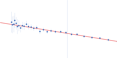
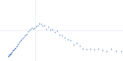
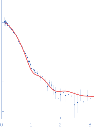
SANS data from solutions of His-tagged L-methionine gamma-lyase from Clostridium tetani in PBS-D2O (137 mM NaCl, 2.7 mM KCl, 10 mM Na2HPO4, 1.8 mM KH2PO4 (D2O buffer), pD 7.4) performed on the YuMO SANS TOF spectrometer (IBR-2: Frank Laboratory of Neutron Physics, Joint Institute for Nuclear Research; Dubna, Russian Federation) with a two He3-fulfilled, 8 independent wire detector system (sample-detector distances of 4.5 m and 12.97 m). The wavelengths used are from 0.05 to 0.8 nm, the contributions of which are separated using time-of-flight technology. One solute concentration of 10.00 mg/ml was measured at 20°C. 9000 successive 0.207 second frames were collected.

The protein of study is His-tagged L methionine gamma lyase from Clostridium tetani. The volume fractions of tetramers and octamers in solution are 37% and 63%, respectively.

L-methionine gamma-lyase (Ct-MGL)
Mol. type   Protein
Organism   Clostridium tetani
Olig. state   Tetramer
Mon. MW   45.4 kDa
 
UniProt   A0A1L7H888 (1-398)
Sequence   FASTA
 
PDB ID   5DX5
 
PDB ID   5DX5