|
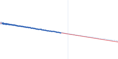
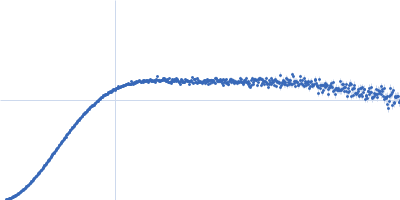
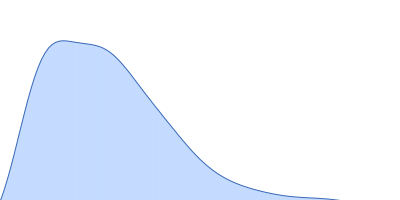
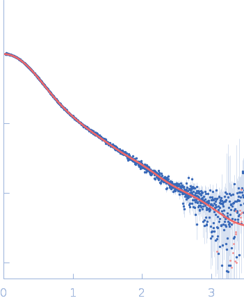
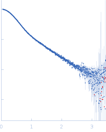
Synchrotron SAXS data from solutions of duplex-G4-duplex DNA in 6 mM Na2HPO4, 2 mM NaH2PO4, 1 mM Na2EDTA, 185 mM KCl, pH 7.2 were collected on the BioCAT 18ID beam line at the Advanced Photon Source (APS), Argonne National Laboratory (Lemont, IL, USA) using a Pilatus3 X 1M detector at a sample-detector distance of 3.6 m and at a wavelength of λ = 0.1033 nm (I(s) vs s, where s = 4πsinθ/λ, and 2θ is the scattering angle). In-line size-exclusion chromatography (SEC) SAS was employed. The SEC parameters were as follows: A 240.00 μl sample at 6.6 mg/ml was injected at a 0.60 ml/min flow rate onto a GE Superdex 75 Increase 10/300 column at 25°C. The data were normalized to the intensity of the transmitted beam and radially averaged; the scattering of the solvent-blank was subtracted.
Sample is a synthetic duplex-G4-duplex DNA with a poly dT surrogate complement loop to facilitate G4 folding and maintenance.
|
|
Duplex-G4-duplex
(DGD)
|
| Mol. type |
|
DNA |
| Organism |
|
synthetic construct |
| Olig. state |
|
Monomer |
| Mon. MW |
|
28.6 kDa |
| Sequence |
|
FASTA |
| |
|
 s, nm-1