Glutathione S-Transferase Mediated Epoxide Conversion: Structural Insights into an Enantioselective Catalyst

Jens Reiners.

SASDQY7 – StyI glutathione S-transferase

GST N-terminal domain-containing protein
MWI(0) 61 kDa
MWexpected 60 kDa
VPorod 91 nm3
log I(s) 4.43×10⁻² 4.43×10⁻³ 4.43×10⁻⁴ 4.43×10⁻⁵
GST N-terminal domain-containing protein small angle scattering data  s, nm-1
ln I(s)
GST N-terminal domain-containing protein Guinier plot ln 4.44×10⁻² Rg: 2.7 nm 0 (2.7 nm)-2 s2
(sRg)2I(s)/I(0)
GST N-terminal domain-containing protein Kratky plot 1.104 0 3 sRg
p(r)
GST N-terminal domain-containing protein pair distance distribution function Rg: 2.7 nm 0 Dmax: 9.2 nm

Data validation


Fits and models


log I(s)
 s, nm-1
GST N-terminal domain-containing protein CORAL model

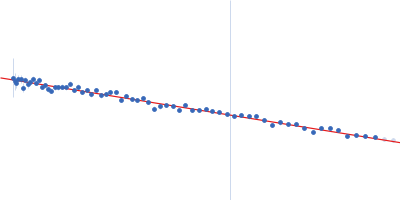
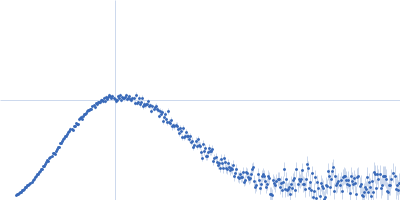
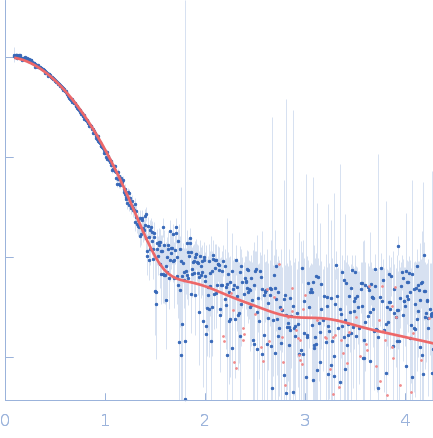
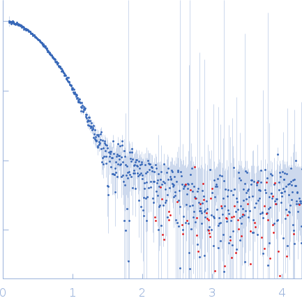
SAXS data from solutions of StyI glutathione S-transferase in 10 mM Tris, 300 mM NaCl, pH 8 were collected using a Xenocs Xeuss 2.0 Q-Zoom instrument equipped with a Pilatus3 R 300 K detector and a GENIX 3D CU Ultra Low Divergence X-ray beam delivery system (Center for Structural Studies, Heinrich-Heine-University, Düsseldorf, Germany) at a sample-detector distance of 0.6 m and at a wavelength of λ = 0.154 nm (I(s) vs s, where s = 4πsinθ/λ, and 2θ is the scattering angle). One solute concentration of 4.70 mg/ml was measured at 10°C. 12 successive 600 second frames were collected. The data were normalized to the intensity of the transmitted beam and radially averaged; the scattering of the solvent-blank was subtracted.

GST N-terminal domain-containing protein (Styl)
Mol. type   Protein
Organism   Gordonia rubripertincta
Olig. state   Dimer
Mon. MW   29.8 kDa
 
UniProt   A0A222TSG8 (1-238)
Sequence   FASTA