Synchrotron SAXS
data from solutions of
Trypsin + Aprotinin: Complex equilibrium
in
20 mM TRIS, 40 mM KCl, 20 mM CaCl2, pH 7
were collected
on the
G1 beam line
at the Cornell High Energy Synchrotron Source (CHESS) storage ring
(Ithaca, NY, USA)
using a Pilatus 100K detector
at a wavelength of λ = 0.10972 nm
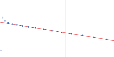
(I(s) vs s, where s = 4πsinθ/λ, and 2θ is the scattering angle).
One solute concentration of 5.33 mg/ml was measured
at 20°C.
20 successive
5 second frames were collected.
The data were normalized to the intensity of the transmitted beam and radially averaged; the scattering of the solvent-blank was subtracted.
Aprotinin (1.73 mg/mL) and trypsin (4 mg/mL) were combined and static data point was taken minutes later. Molecular weight determined with Vc
 s, nm-1
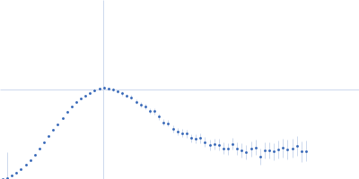