|
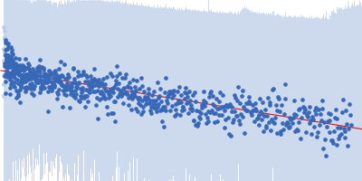
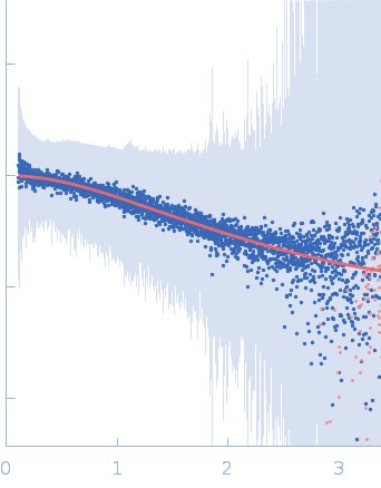
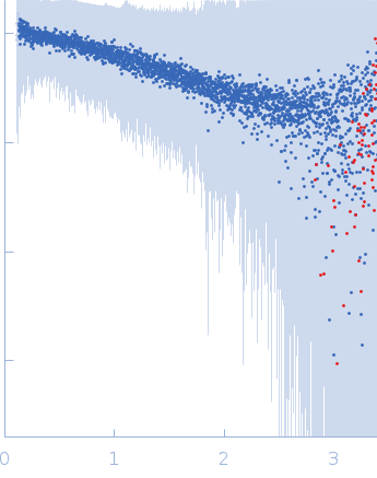
Synchrotron SAXS
data from solutions of
Segment S(26-45) of the Neurofilament low intrinsically disordered tail domain
in
20 mM Tris, pH 8
were collected
on the
B21 beam line
at the Diamond Light Source storage ring
(Didcot, UK)
using a Eiger 4M detector
at a wavelength of λ = 0.1 nm
(I(s) vs s, where s = 4πsinθ/λ, and 2θ is the scattering angle).
One solute concentration of 1.00 mg/ml was measured
at 24°C.
The data were normalized to the intensity of the transmitted beam and radially averaged; the scattering of the solvent-blank was subtracted.
Sample detector distance = UNKNOWN. Number of frames = UNKNOWN
|
|
 s, nm-1
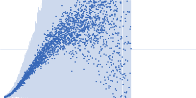
 of the Neurofilament low intrinsically disordered tail domain Rg histogram") Rg, nm
Rg, nm