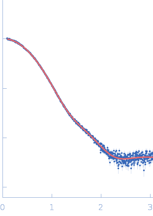
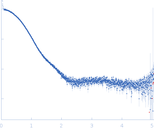
Sugar binding to Kluyveromyces lactis glucokinase 1 (KlGlk1)

Renato H. Weiße.

SASDUD6 – Extrapolated scattering curve of monomeric Kluyveromyces lactis glucokinase 1 triple point mutant I356D, Y419D, H420D

Glucokinase-1 (I356D, Y419D, H420D)
MWexperimental 51 kDa
MWexpected 54 kDa
VPorod 78 nm3
log I(s) 3.36×10⁻² 3.36×10⁻³ 3.36×10⁻⁴ 3.36×10⁻⁵
Glucokinase-1 (I356D, Y419D, H420D) small angle scattering data  s, nm-1
ln I(s)
Glucokinase-1 (I356D, Y419D, H420D) Guinier plot ln 3.37×10⁻² Rg: 2.6 nm 0 (2.6 nm)-2 s2
(sRg)2I(s)/I(0)
Glucokinase-1 (I356D, Y419D, H420D) Kratky plot 1.104 0 3 sRg
p(r)
Glucokinase-1 (I356D, Y419D, H420D) pair distance distribution function Rg: 2.6 nm 0 Dmax: 9 nm

Data validation


Fits and models


log I(s)
 s, nm-1
Glucokinase-1 (I356D, Y419D, H420D) DAMMIN model
Glucokinase-1 (I356D, Y419D, H420D) PYMOL model

Synchrotron SAXS data from solutions of Extrapolated scattering curve of monomeric Kluyveromyces lactis glucokinase 1 triple point mutant I356D, Y419D, H420D in 10 mM Tris-HCl, 1 mM DTT, pH 7.4 were collected on the EMBL P12 beam line at the PETRA III storage ring (DESY; Hamburg, Germany) using a Pilatus 2M detector at a sample-detector distance of 3 m and at a wavelength of λ = 0.1239 nm (I(s) vs s, where s = 4πsinθ/λ, and 2θ is the scattering angle). Solute concentrations ranging between 10 and 3 mg/ml were measured at 10°C. 30 successive 0.045 second frames were collected. The data were normalized to the intensity of the transmitted beam and radially averaged; the scattering of the solvent-blank was subtracted. The low angle data collected at lower concentrations were extrapolated to infinite dilution and merged with the higher concentration data to yield the final composite scattering curve.

Glucokinase-1 (I356D, Y419D, H420D) (KlGlk1_mon)
Mol. type   Protein
Organism   Kluyveromyces lactis (strain ATCC 8585 / CBS 2359 / DSM 70799 / NBRC 1267 / NRRL Y-1140 / WM37)
Olig. state   Monomer
Mon. MW   53.7 kDa
 
UniProt   Q6CUZ3 (1-481)
Sequence   FASTA