Sugar binding to Kluyveromyces lactis glucokinase 1 (KlGlk1)

Renato H. Weiße.

SASDUE6 – Monomeric Kluyveromyces lactis glucokinase 1 triple point mutant (I356D, Y419D, H420D), 3.0 mg/mL, no ligand

Glucokinase-1 (I356D, Y419D, H420D)
MWexperimental 46 kDa
MWexpected 54 kDa
VPorod 76 nm3
log I(s) 2.90×10⁻² 2.90×10⁻³ 2.90×10⁻⁴ 2.90×10⁻⁵
Glucokinase-1 (I356D, Y419D, H420D) small angle scattering data  s, nm-1
ln I(s)
Glucokinase-1 (I356D, Y419D, H420D) Guinier plot ln 2.90×10⁻² Rg: 2.4 nm 0 (2.4 nm)-2 s2
(sRg)2I(s)/I(0)
Glucokinase-1 (I356D, Y419D, H420D) Kratky plot 1.104 0 3 sRg
Dmax: 8 nm

Data validation


Fits and models


log I(s)
 s, nm-1
Glucokinase-1 (I356D, Y419D, H420D) PYMOL model

Synchrotron SAXS data from solutions of Monomeric Kluyveromyces lactis glucokinase 1 triple point mutant (I356D, Y419D, H420D), 3.0 mg/mL, no ligand in 10 mM Tris-HCl, 1 mM DTT, pH 7.4 were collected on the EMBL P12 beam line at the PETRA III storage ring (DESY; Hamburg, Germany) using a Pilatus 2M detector at a sample-detector distance of 3 m and at a wavelength of λ = 0.1239 nm 
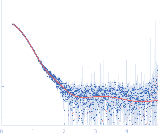
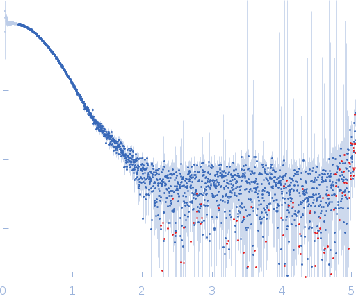
(I(s) vs s, where s = 4πsinθ/λ, and 2θ is the scattering angle). Solute concentrations ranging between 14.2 and 3 mg/ml were measured at 10°C. 30 successive 0.045 second frames were collected. The data were normalized to the intensity of the transmitted beam and radially averaged; the scattering of the solvent-blank was subtracted.

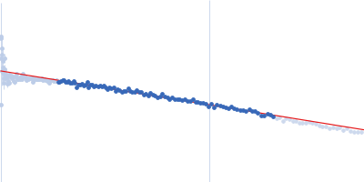
CAUTION: Repulsive interparticle interference effects present in the sample result in inaccurate structural parameters (Rg, I(0), MW, Dmax) extracted from the data.

Glucokinase-1 (I356D, Y419D, H420D) (KlGlk1_mon)
Mol. type   Protein
Organism   Kluyveromyces lactis (strain ATCC 8585 / CBS 2359 / DSM 70799 / NBRC 1267 / NRRL Y-1140 / WM37)
Olig. state   Monomer
Mon. MW   53.7 kDa
 
UniProt   Q6CUZ3 (1-481)
Sequence   FASTA