Solution Structure Determination and Biophysical Studies of 7SK RNP and 7SL SRP RNAs.

D'Souza MH, Pereira HS, Tersteeg S, Vasudeva G, Siddiqui MQ, Agunlejika T, Kerr L, Girodat D, Patel T, RNA (2025) Europe PMC

SASDVE9 – 7SL Signal Recognition Particle

7SL Signal Reocgnition Particle RNA
MWexperimental 98 kDa
MWexpected 98 kDa
VPorod 300 nm3
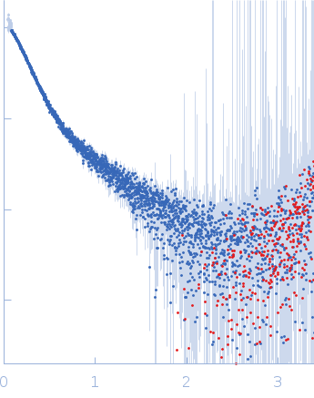
log I(s) 3.63×10⁻² 3.63×10⁻³ 3.63×10⁻⁴ 3.63×10⁻⁵
7SL Signal Reocgnition Particle RNA small angle scattering data  s, nm-1
ln I(s)
7SL Signal Reocgnition Particle RNA Guinier plot ln 3.63×10⁻² Rg: 6.7 nm 0 (6.7 nm)-2 s2
(sRg)2I(s)/I(0)
7SL Signal Reocgnition Particle RNA Kratky plot 1.104 0 3 sRg
p(r)
7SL Signal Reocgnition Particle RNA pair distance distribution function Rg: 6.5 nm 0 Dmax: 21.7 nm

Data validation


Fits and models


log I(s)
 s, nm-1
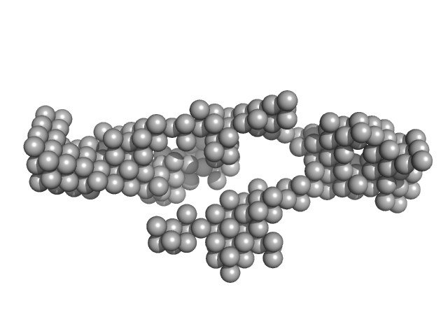
7SL Signal Reocgnition Particle RNA DAMMIN model

log I(s)
 s, nm-1
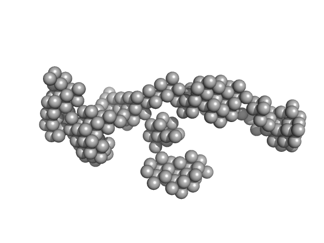
7SL Signal Reocgnition Particle RNA DAMMIN model

log I(s)
 s, nm-1
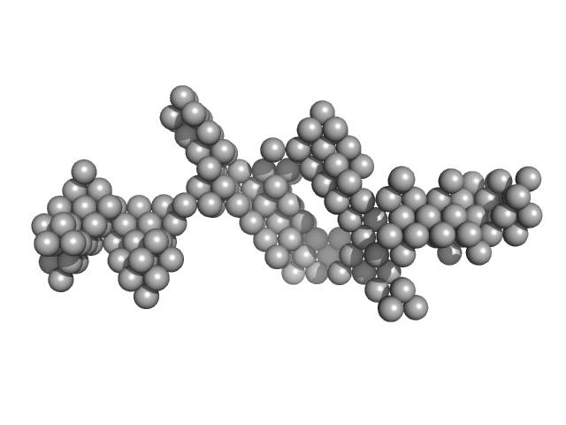
7SL Signal Reocgnition Particle RNA DAMMIN model

Synchrotron SAXS data from solutions of 7SL Signal Recognition Particle in 10 mM Bis-Tris, 100 mM NaCl, 15 mM KCl, 5% Glycerol, 3 mM MgCl2, pH 5.5 were collected on the B21 beam line at the Diamond Light Source storage ring (Didcot, UK) using a Eiger 4M detector at a sample-detector distance of 3.7 m and at a wavelength of λ = 0.094 nm (I(s) vs s, where s = 4πsinθ/λ, and 2θ is the scattering angle). One solute concentration of 1.00 mg/ml was measured at 20°C. 600 successive 3 second frames were collected. The data were normalized to the intensity of the transmitted beam and radially averaged; the scattering of the solvent-blank was subtracted.

7SL Signal Reocgnition Particle RNA (7SL)
Mol. type   RNA
Organism   Homo sapiens
Olig. state   Monomer
Mon. MW   98.2 kDa
Sequence   FASTA