|
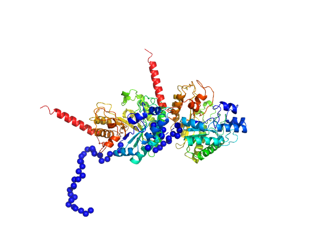
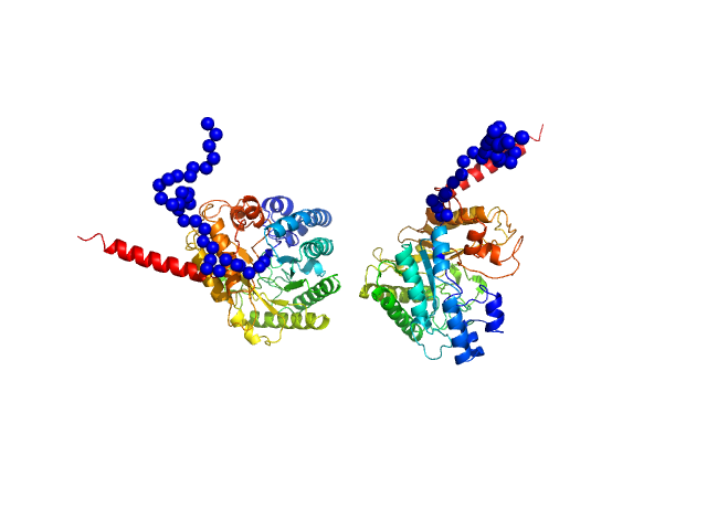
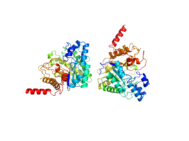
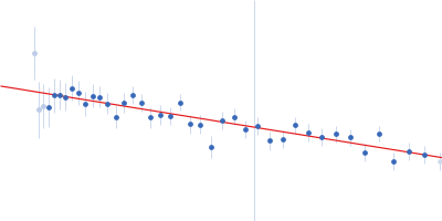
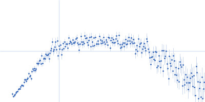
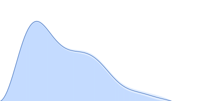
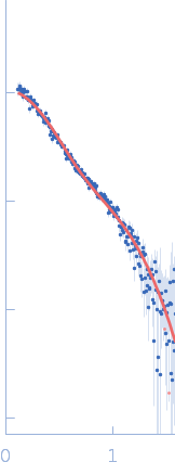
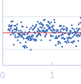
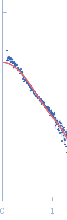
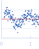
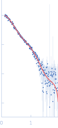
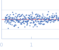
SAXS data from solutions of Glycosidase/Rutinosidase (deglycosylated dimer) in 50 mM sodium citrate, 150 mM NaCl, pH 4.5 were collected using an Anton Paar SAXSpoint 2.0 instrument at the Institute of Biotechnology, Czech Academy of Sciences/Centre of Molecular Structure (Vestec, Czech Republic) equipped with an Eiger R 1M detector at a sample-detector distance of 0.8 m and at a wavelength of λ = 0.134 nm (I(s) vs s, where s = 4πsinθ/λ, and 2θ is the scattering angle). Solute concentrations ranging between 0.7 and 0.9 mg/ml were measured at 20°C. 40 successive 15 second frames were collected. The data were normalized to the intensity of the transmitted beam and radially averaged; the scattering of the solvent-blank was subtracted. The low angle data collected at lower concentration were merged with the highest concentration high angle data to yield the final composite scattering curve.
Glycosidase/Rutinosidase from Mucor circinelloides was treated using endo H glycosidase. Two runs of SEC-SAXS with different injection concentration were performed. The resulting data buffer subtracted data from the peaks were merged to mitigate the repulsive inter-particle interaction.
The raw data are available at: https://doi.org/10.5281/zenodo.13946174
|
|

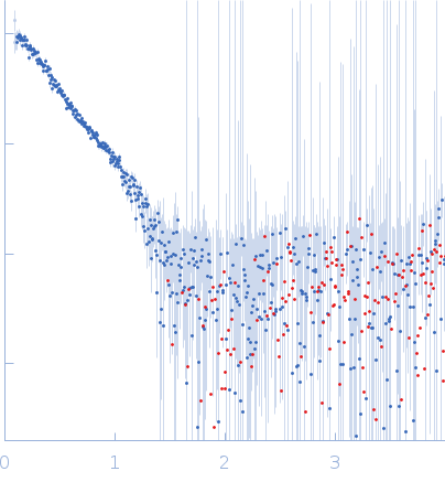 s, nm-1
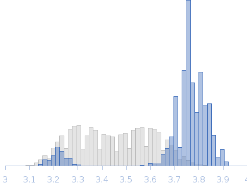 Rg, nm