|
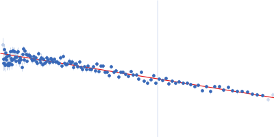
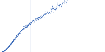
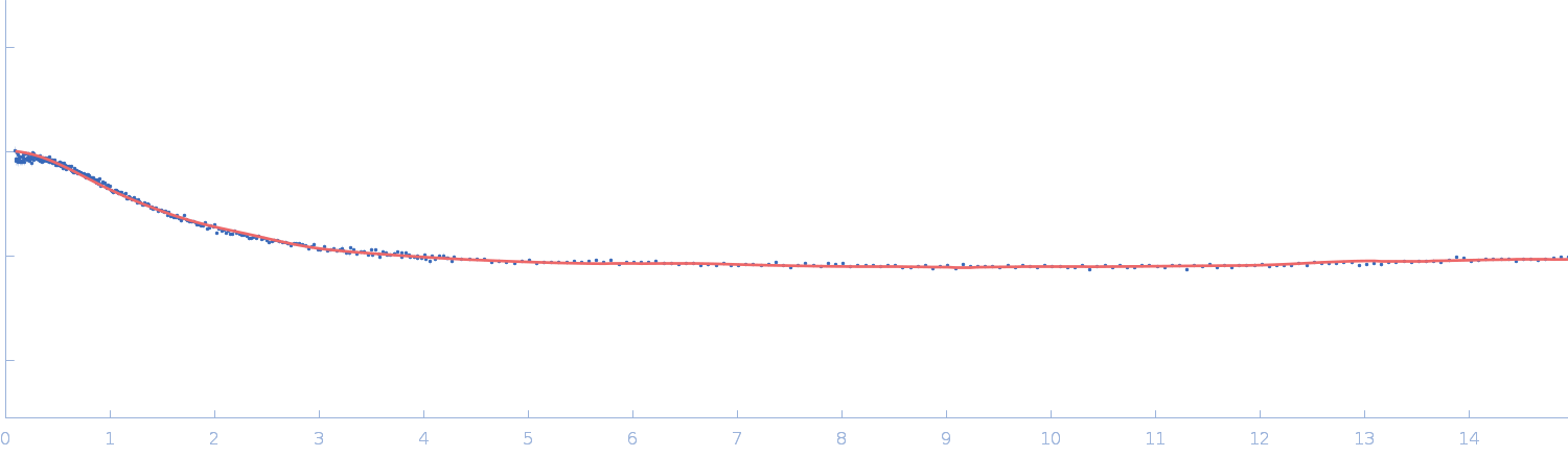
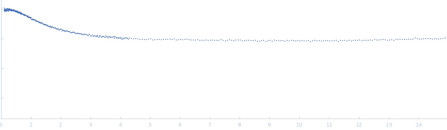
Synchrotron SAXS data from solutions of HTT0 peptide in sodium phosphate buffer, pH 7.4 were collected on the TPS13A beam line at the NSRRC (Hsinchu, Taiwan) using a Eiger X 1M & 9M detector at a sample-detector distance of 2.5 m and at a wavelength of λ = 0.08265 nm (I(s) vs s, where s = 4πsinθ/λ, and 2θ is the scattering angle). In-line size-exclusion chromatography (SEC) SAS was employed. The SEC parameters were as follows: A 70.00 μl sample at 10 mg/ml was injected at a 0.35 ml/min flow rate onto a Agilent Bio SEC-3, 300 Å column at 10°C. 10 successive 2 second frames were collected. The data were normalized to the intensity of the transmitted beam and radially averaged; the scattering of the solvent-blank was subtracted.
The sodium phosphate buffer solution contains 480 μL of 10 mM PB, pH 7.4, 16.5 μL of 100 mM NaOH, and 10 μL of 1% trifluoroacetic acid (TFA).
|
|
HTT0
|
| Mol. type |
|
Protein |
| Organism |
|
synthetic construct |
| Olig. state |
|
Monomer |
| Mon. MW |
|
4.0 kDa |
| Sequence |
|
FASTA |
| |
|
 s, nm-1