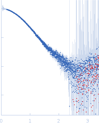
Nanobodies as tools for studying human frataxin biology.

Pignataro MF, Fernández NB, Garay-Alvarez A, Pavan MF, Molina R, Muñoz IG, Grossi J, Noguera M, Vila A, García AE, Gentili HG, Rodríguez NA, Aran M, Parreño V, Bok M, Hermoso JA, Ibañez LI, Santos J, Commun Biol (2026) Europe PMC

SASDWS6 – Frataxin bound to tailored nanobody 28F6

Frataxin, mitochondrial
Nanobody 28F6
MWI(0) 25 kDa
MWexpected 29 kDa
VPorod 36 nm3
log I(s) 3.32×10⁻² 3.32×10⁻³ 3.32×10⁻⁴ 3.32×10⁻⁵
Frataxin, mitochondrial Nanobody 28F6 small angle scattering data  s, nm-1
ln I(s)
Frataxin, mitochondrial Nanobody 28F6 Guinier plot ln 3.32×10⁻² Rg: 2.2 nm 0 (2.2 nm)-2 s2
(sRg)2I(s)/I(0)
Frataxin, mitochondrial Nanobody 28F6 Kratky plot 1.104 0 3 sRg
p(r)
Frataxin, mitochondrial Nanobody 28F6 pair distance distribution function Rg: 2.3 nm 0 Dmax: 7.4 nm

Data validation


Fits and models


log I(s)
 s, nm-1
Frataxin, mitochondrial Nanobody 28F6 ALPHAFOLD model

log I(s)
 s, nm-1
Frataxin, mitochondrial Nanobody 28F6 DAMMIN model

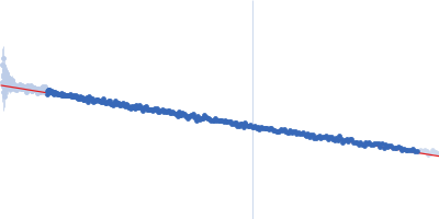
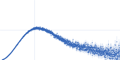
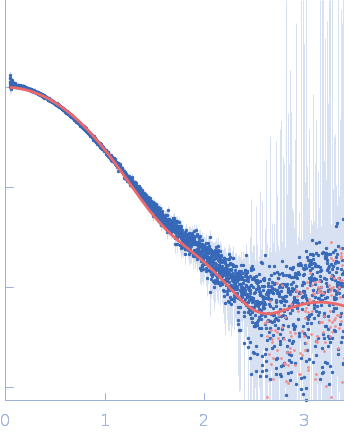
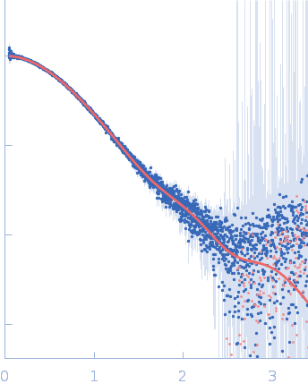
Synchrotron SAXS data from solutions of Frataxin bound to tailored nanobody 28F6 in 20 mM Tris, 150 mM NaCl, pH 7.5 were collected on the B21 beam line at the Diamond Light Source (Didcot, UK) using a Eiger 4M detector at a sample-detector distance of 3.7 m and at a wavelength of λ = 0.1 nm (I(s) vs s, where s = 4πsinθ/λ, and 2θ is the scattering angle). One solute concentration of 4.00 mg/ml was measured at 18°C. 600 successive 3 second frames were collected. The data were normalized to the intensity of the transmitted beam and radially averaged; the scattering of the solvent-blank was subtracted.

Frataxin, mitochondrial (FXN)
Mol. type   Protein
Organism   Homo sapiens
Olig. state   Monomer
Mon. MW   13.6 kDa
 
UniProt   Q16595 (90-210)
Sequence   FASTA
 
Nanobody 28F6 (28F6)
Mol. type   Protein
Organism   synthetic construct
Olig. state   Monomer
Mon. MW   15.3 kDa
Sequence   FASTA