Targeting the phosphoprotein enriched in diabetes/astrocytes 15 death effector domain via fragment-based and integrative structural biology approaches.

Mercurio I, Carucci F, Nanna V, Di Marino S, Papadopoulos MGE, Bischetti M, Fata F, Malgieri G, Siliqi D Piai A, Fattorusso R, Mangiatordi GF, Toniatti C, Saviano M, Alberga D, Int J Biol Macromol :154849 (2026) Europe PMC

SASDX35 – DD mutant astrocytic phosphoprotein PEA-15

Astrocytic phosphoprotein PEA-15 (S104D, S116D)
MWexperimental 15 kDa
MWexpected 15 kDa
VPorod 27 nm3
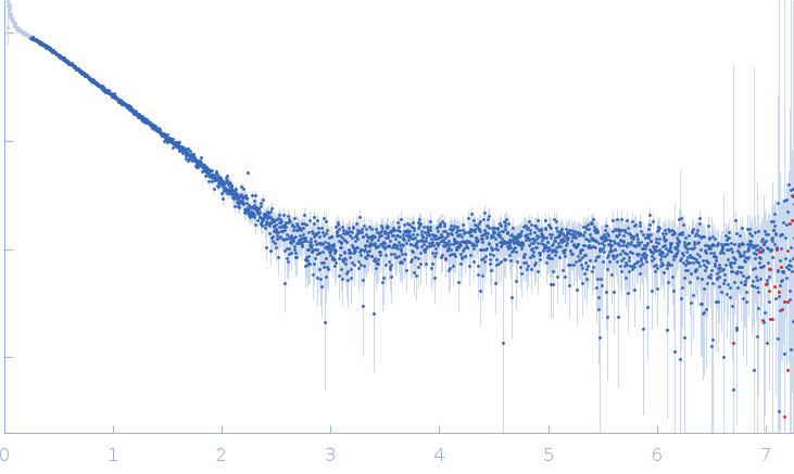
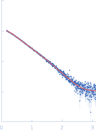
log I(s) 2.39×10⁻² 2.39×10⁻³ 2.39×10⁻⁴ 2.39×10⁻⁵
Astrocytic phosphoprotein PEA-15 (S104D, S116D) small angle scattering data  s, nm-1
ln I(s)
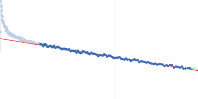
Astrocytic phosphoprotein PEA-15 (S104D, S116D) Guinier plot ln 2.39×10⁻² Rg: 2.4 nm 0 (2.4 nm)-2 s2
(sRg)2I(s)/I(0)
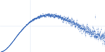
Astrocytic phosphoprotein PEA-15 (S104D, S116D) Kratky plot 1.104 0 √3 sRg
p(r)
Astrocytic phosphoprotein PEA-15 (S104D, S116D) pair distance distribution function Rg: 2.9 nm 0 Dmax: 12.7 nm

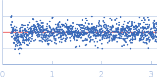
Data validation


Fits and models


log I(s)
 s, nm-1
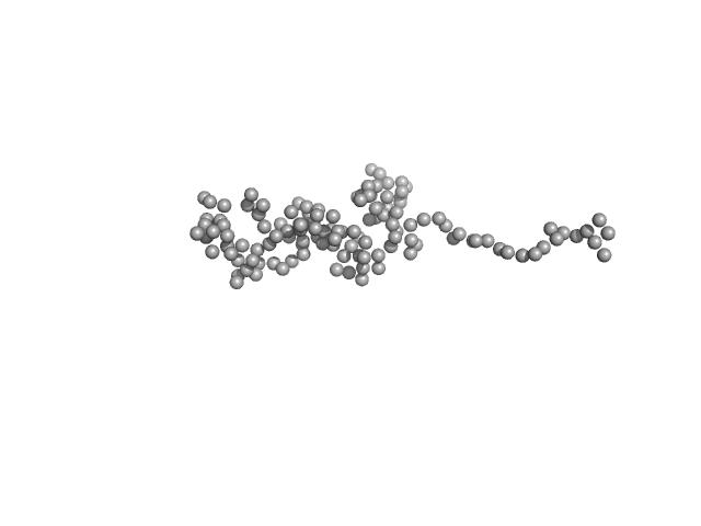
Astrocytic phosphoprotein PEA-15 (S104D, S116D) GASBOR model

log I(s)
 s, nm-1
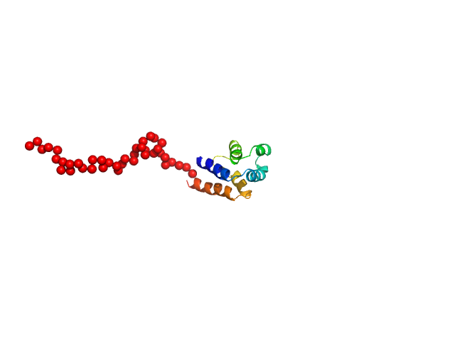
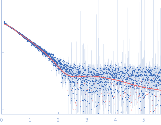
Astrocytic phosphoprotein PEA-15 (S104D, S116D) CORAL model

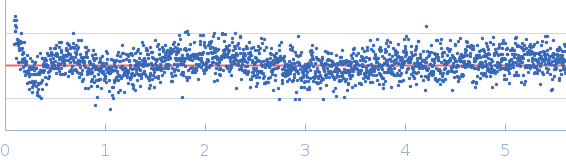
Synchrotron SAXS data from solutions of PEA15-DD mutant, in 20 mM Na2PO4 2mM DTT pH=7.0 were collected on the P12 beamline at DESY (Hamburg, Germany) using a Pilatus 6m detector at a sample-detector distance of 3.0 m and a wavelength of λ = 0.124 nm (I(s) vs s, where s = 4πsinθ/λ, and 2θ is the scattering angle). A solute concentration of 0.6 to 2.2 mg/ml was measured at 20°C. 20 successive 0.1-second frames were collected. The data were normalized to the intensity of the transmitted beam and radially averaged and brought into the absolute scale with respect to the intensity of the empty capillary and the water; the scattering of the solvent-blank was subtracted. First, a Ca model was reconstructed by GASBOR which fits quite well with the NMR model (PDB ID 1N3K). Second, the long C-tail was added to the NMR model as dummy-residues, where the spatial position of the tail was optimised using the program CORAL.The goal of SAXS data was to assess differences in the spatial sampling/conformation of the DD mutant variant compared to the wild-type protein.

Astrocytic phosphoprotein PEA-15 (S104D, S116D) (DD PEA15)
Mol. type   Protein
Organism   Cricetulus griseus
Olig. state   Monomer
Mon. MW   15.1 kDa
 
UniProt   Q9Z297 (1-130)
Sequence   FASTA
 
PDB ID   1N3K