|
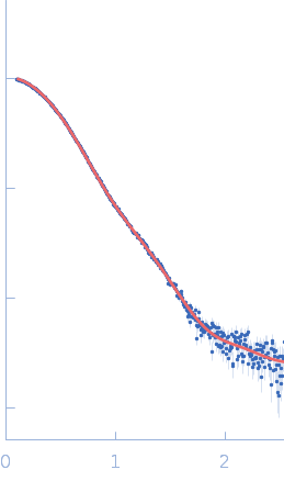
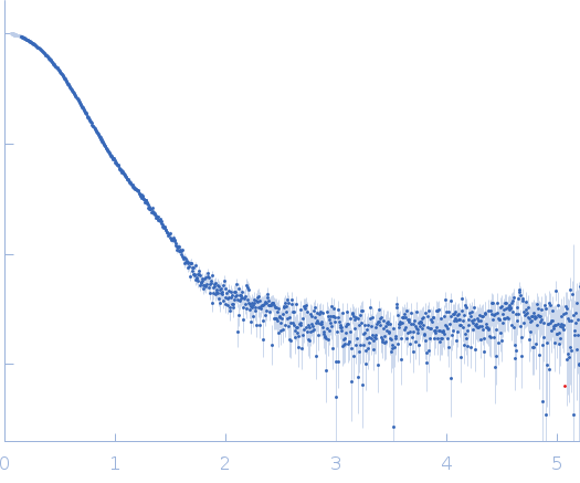
Synchrotron SAXS
data from solutions of
Rv2514c-Rv2515c VapBC52 complex
in
20 mM HEPES, 150 mM NaCl, 10% glycerol, pH 8
were collected
on the
BM29 beam line
at the ESRF storage ring
(Grenoble, France)
using a Pilatus3 2M detector
at a sample-detector distance of 2.8 m and
at a wavelength of λ = 0.099 nm
(I(s) vs s, where s = 4πsinθ/λ, and 2θ is the scattering angle).
One solute concentration of 1.00 mg/ml was measured
at 20°C.
700 successive
2 second frames were collected.
The data were normalized to the intensity of the transmitted beam and radially averaged; the scattering of the solvent-blank was subtracted.
|
|
Ribonuclease VapC52
(VapC52)
|
| Mol. type |
|
Protein |
| Organism |
|
Mycobacterium tuberculosis |
| Olig. state |
|
Dimer |
| Mon. MW |
|
18.6 kDa |
| Sequence |
|
FASTA |
| |
|
Antitoxin VapB52
(VapB52)
|
| Mol. type |
|
Protein |
| Organism |
|
Mycobacterium tuberculosis |
| Olig. state |
|
Monomer |
| Mon. MW |
|
43.3 kDa |
| Sequence |
|
FASTA |
| |
|
 s, nm-1