Synchrotron SAXS
data from solutions of
Phosphorylated SARS-CoV-2 Nucleocapsid protein 1.5 mg/mL
in
20 mM Tris, 150 mM NaCl, pH 7.5
were collected
on the
16-ID (LiX) beam line
at the National Synchrotron Light Source II (NSLS-II) storage ring
(Upton, NY, USA)
using a Pilatus3 S 1M, Pilatus3 900 K detector
at a sample-detector distance of 3.6 m and
at a wavelength of λ = 0.08189 nm
(I(s) vs s, where s = 4πsinθ/λ, and 2θ is the scattering angle).
One solute concentration of 1.50 mg/ml was measured
at 25°C.
10 successive
0.500 second frames were collected.
The data were normalized to the intensity of the transmitted beam and radially averaged; the scattering of the solvent-blank was subtracted.
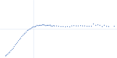
Phosphorylated SARS-Cov-2 Nucleocapsid protein dimers at 1.5 mg/mL concentration in 150mM NaCl, 20mM Tris.
Phosphorylation carried out in vitro using GSK3b and SRPK1 kinases.
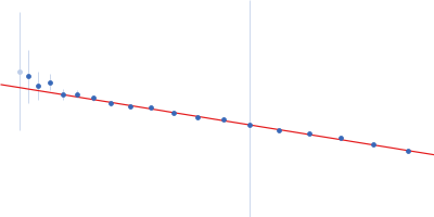
SAXS data was collected over a s range of 0.05 - 31.3 nm-1 and analyzed within a range of 0.05 - 2.55 nm-1.
λ = 0.08189 nm. Sample to detector distance for small angles was 3.560 m and 0.317 m for wide angles using Pilatus3X 1M and Pilatus3X 900k detectors.
Background subtraction was performed for each profile from its buffer.
Data collected at NSLS2 at Upton, NY.
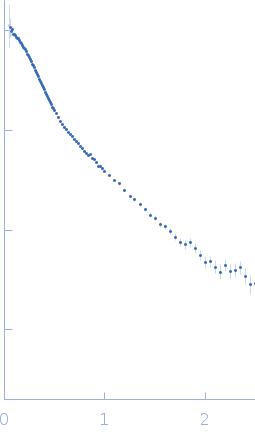 s, nm-1