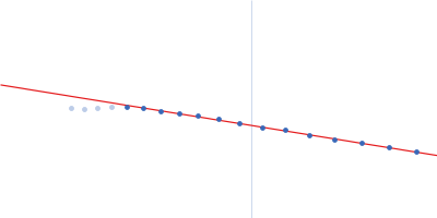
| MWexperimental | 105 | kDa |
| MWexpected | 100 | kDa |
| VPorod | 154 | nm3 |
|
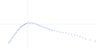
log I(s)
4.90×10⁻²
4.90×10⁻³
4.90×10⁻⁴
4.90×10⁻⁵
|
 s, nm-1
|
|
|
|

|
|

|
|

|
|
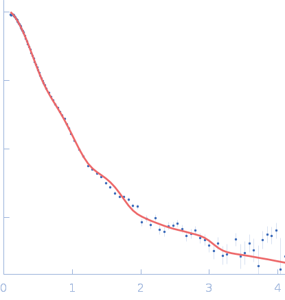
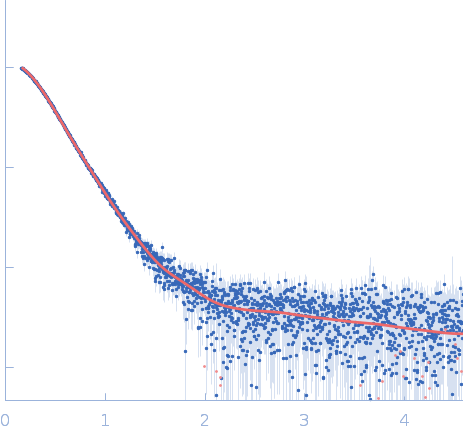
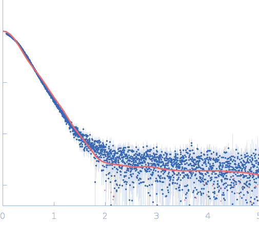
Synchrotron SAXS
data from solutions of
RTX cytotoxin pro-MbxA without acylation (pro-MbxA)
in
50 mM Tris, 100 mM NaCl, 10mM CaCl2, pH 7.8
were collected
on the
EMBL P12 beam line
at the PETRA III storage ring
(DESY; Hamburg, Germany)
using a Pilatus 6M detector
at a sample-detector distance of 3 m and
at a wavelength of λ = 0.155 nm
(I(s) vs s, where s = 4πsinθ/λ, and 2θ is the scattering angle).
In-line size-exclusion chromatography (SEC) SAS was employed. The SEC parameters were as follows: A 100.00 μl sample
at 5.3 mg/ml was injected at a 0.50 ml/min flow rate
onto a GE Superose 6 Increase 10/300 column
at 20°C.
3000 successive
0.995 second frames were collected.
The data were normalized to the intensity of the transmitted beam and radially averaged; the scattering of the solvent-blank was subtracted.
|
|
|||||||||||||||||||||||||||