Bacterial targeting of the neutrophil inhibitory receptor LILRB3 to evade antibody immunity

Ying Zhang.

SASDXH8 – Leukocyte Immunoglobulin-Like Receptor LILR-B3

Leukocyte immunoglobulin-like receptor subfamily B member 3 (P288R)
MWexperimental 58 kDa
MWexpected 47 kDa
VPorod 137 nm3
log I(s) 2.75×101 2.75×100 2.75×10⁻¹ 2.75×10⁻²
Leukocyte immunoglobulin-like receptor subfamily B member 3 (P288R) small angle scattering data  s, nm-1
ln I(s)
Leukocyte immunoglobulin-like receptor subfamily B member 3 (P288R) Guinier plot ln 2.76×101 Rg: 3.6 nm 0 (3.6 nm)-2 s2
(sRg)2I(s)/I(0)
Leukocyte immunoglobulin-like receptor subfamily B member 3 (P288R) Kratky plot 1.104 0 3 sRg
p(r)
Leukocyte immunoglobulin-like receptor subfamily B member 3 (P288R) pair distance distribution function Rg: 3.8 nm 0 Dmax: 13.5 nm

Data validation


Fits and models


log I(s)
 s, nm-1
Leukocyte immunoglobulin-like receptor subfamily B member 3 (P288R) MODELLER model

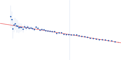
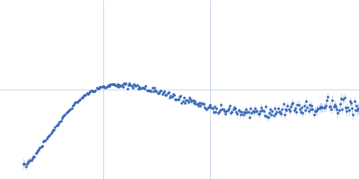
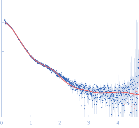
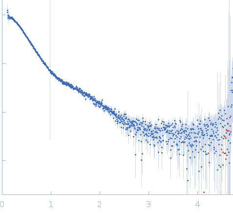
Synchrotron small-angle X-ray scattering (SAXS) data on Leukocyte Immunoglobulin-Like Receptor (LILR-B3) were collected at SIBYLS beamline 12.3.1 of the Advanced Light Source in SEC-SAXS mode. In-line size-exclusion chromatography (SEC) was performed using an Agilent 1260 HPLC system. The SEC conditions were as follows: a 60 µL sample at 5 mg/mL was injected onto a Shodex KW-803 column at 4 °C that had been previously equilibrated in 20 mM HEPES, 140 mM NaCl, pH 7.4, and eluted at 0.65 mL/min. Scattering was recorded on a Pilatus3 X 2M detector at a sample-to-detector distance of 2.1 m and a wavelength of λ = 0.1127 nm (I(q) vs q; q = 4π sin θ/λ, where 2θ is the scattering angle). 913 successive 2-s frames were collected. The scattering curves were normalized to the transmitted-beam intensity, radially averaged, and corrected by subtracting the solvent blank.

Leukocyte immunoglobulin-like receptor subfamily B member 3 (P288R) (LILRB3)
Mol. type   Protein
Organism   Homo sapiens
Olig. state   Monomer
Mon. MW   47.0 kDa
 
UniProt   A0A0G2JM46 (24-443)
Sequence   FASTA