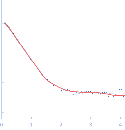
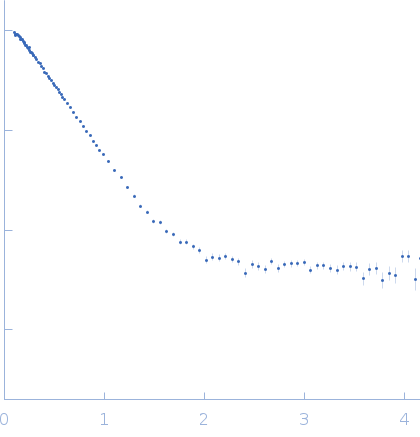
Dynamics and structural features of the eEF1A1 and eEF1A2 paralogs

Novosylna O, Shalak V, Dąbrowska K, Patmanidis I, Lozhko D Bondarchuk T, Schiøtt B, Pedersen J, Knudsen C, Nissen P, Dadlez M, Negrutskii B, Nucleic Acids Research 53(21) (2025) DOI

SASDXJ2 – Mammalian translation elongation factor eEF1A1

Mammalian translation elongation factor eEF1A1
MWI(0) 134 kDa
MWexpected 100 kDa
VPorod 144 nm3
log I(s) 1.51×10⁻¹ 1.51×10⁻² 1.51×10⁻³ 1.51×10⁻⁴
Mammalian translation elongation factor eEF1A1 small angle scattering data  s, nm-1
ln I(s)
Mammalian translation elongation factor eEF1A1 Guinier plot ln 1.52×10⁻¹ Rg: 4.6 nm 0 (4.6 nm)-2 s2
(sRg)2I(s)/I(0)
Mammalian translation elongation factor eEF1A1 Kratky plot 1.104 0 3 sRg
p(r)
Mammalian translation elongation factor eEF1A1 pair distance distribution function Rg: 44.2 nm 0 Dmax: 143.1 nm

Data validation


Fits and models


log I(s)
 s, nm-1
Mammalian translation elongation factor eEF1A1 CHIMERA model

SAXS data from solutions of Mammalian translation elongation factor eEF1A1 in 25 mM Tris HCl, 150 mM NaCl, 6 mM βME, 20% glycerol, 0.01mM GDP,, pH 7.5 were collected on the Bruker Nanostar w Excillum source instrument (Department of Chemistry, iNANO building, Aarhus Uinversity, Aarhus C, Denmark) using a VÅNTEC-2000 detector at a sample-detector distance of 0.9 m and at a wavelength of λ = 0.134 nm (I(s) vs s, where s = 4πsinθ/λ, and 2θ is the scattering angle). One solute concentration of 1.70 mg/ml was measured at 20°C. One 1800 second frame was collected. The data were normalized to the intensity of the transmitted beam and radially averaged; the scattering of the solvent-blank was subtracted.

Mammalian translation elongation factor eEF1A1
Mol. type   Protein
Olig. state   Dimer
Mon. MW   50.1 kDa
 
UniProt   P68104 (1-462)
Sequence   FASTA