Ion-selective conformational stabilization of a disordered repeats-in-toxin protein domain.

Gudinas AP, Shambharkar GM, Chang MP, Fernández D, Matsui T, Mai DJ, Biophys J (2025) Europe PMC

SASDXV8 – Repeats-in-toxin domain Block Vof Bifunctional hemolysin/adenylate cyclase, 1 mM BaCl2

Repeats-in-toxin domain Block V of adenylate cyclase toxin
MWexperimental 21 kDa
MWexpected 19 kDa
VPorod 32 nm3
log I(s) 1.17×10² 1.17×101 1.17×100 1.17×10⁻¹
Repeats-in-toxin domain Block V of adenylate cyclase toxin small angle scattering data  s, nm-1
ln I(s)
Repeats-in-toxin domain Block V of adenylate cyclase toxin Guinier plot ln 1.18×10² Rg: 3.0 nm 0 (3.0 nm)-2 s2
(sRg)2I(s)/I(0)
Repeats-in-toxin domain Block V of adenylate cyclase toxin Kratky plot 1.104 0 3 sRg
p(r)
Repeats-in-toxin domain Block V of adenylate cyclase toxin pair distance distribution function Rg: 3.2 nm 0 Dmax: 13.1 nm

Data validation


Fits and models


log I(s)
 s, nm-1

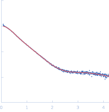
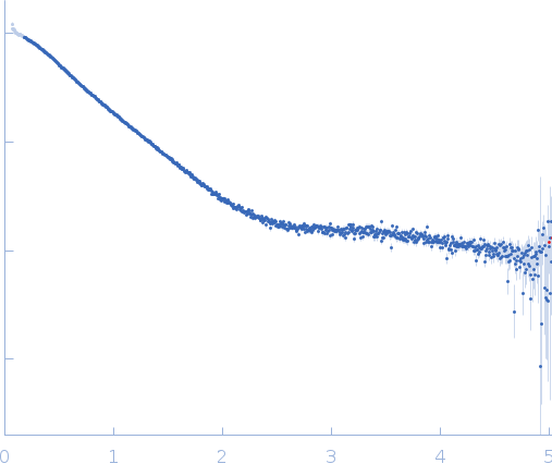
Synchrotron SAXS data from solutions of Disordered repeats-in-toxin domain Block V of Bifunctional hemolysin/adenylate cyclase in 50 mM Tris, 5 mM DTT, pH 7.5 were collected on the BL4-2 beam line at the Stanford Synchrotron Radiation Lightsource (SSRL) storage ring (Menlo Park, CA, USA) using a Pilatus3 X 1M detector at a sample-detector distance of 1.7 m and at a wavelength of λ = 0.1127 nm (I(s) vs s, where s = 4πsinθ/λ, and 2θ is the scattering angle). One solute concentration of 10.00 mg/ml was measured at 21.9°C. 500 frames were collected with an exposure time of 2 seconds. The data were normalized to the intensity of the transmitted beam and radially averaged; the scattering of the solvent blank was subtracted.

Repeats-in-toxin domain Block V of adenylate cyclase toxin (RTX Block V)
Mol. type   Protein
Organism   Bordetella pertussis
Olig. state   Monomer
Mon. MW   19.1 kDa
 
UniProt   P0DKX7 (1529-1680)
Sequence   FASTA