A structure-based epitope tagging approach identifies vulnerable sites on the malarial P36-P52 protein complex for antibody-mediated neutralization of Plasmodium sporozoites.

Das S, Boeykens L, Loubens M, Marinach C, Briquet S, De Vocht L, Pintelon I, Timmermans JP, Sterckx YG Silvie O, PLoS Pathog 22(7):e1014418 (2026) Europe PMC

SASDY36 – Plasmodium falciparum P52-P36 fusion protein

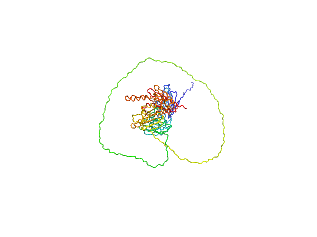
Plasmodium falciparum P52-P36 fusion protein
MWexperimental 83 kDa
MWexpected 84 kDa
VPorod 180 nm3
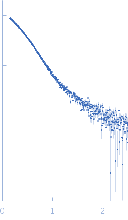
log I(s) 8.45×10⁻¹ 8.45×10⁻² 8.45×10⁻³ 8.45×10⁻⁴
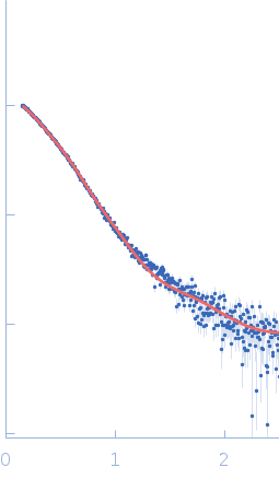
Plasmodium falciparum P52-P36 fusion protein small angle scattering data  s, nm-1
ln I(s)
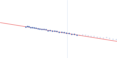
Plasmodium falciparum P52-P36 fusion protein Guinier plot ln 8.46×10⁻¹ Rg: 4.1 nm 0 (4.1 nm)-2 s2
(sRg)2I(s)/I(0)
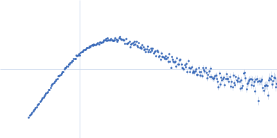
Plasmodium falciparum P52-P36 fusion protein Kratky plot 1.104 0 3 sRg
p(r)
Plasmodium falciparum P52-P36 fusion protein pair distance distribution function Rg: 4.6 nm 0 Dmax: 20.8 nm

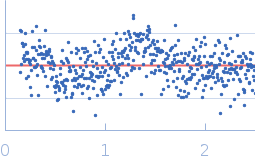
Data validation


Fits and models


log I(s)
 s, nm-1
Plasmodium falciparum P52-P36 fusion protein BILBOMD model

Synchrotron SAXS data from solutions of Plasmodium falciparum P52-P36 fusion protein in 20 mM HEPES, 200 mM NaCl, 3% glycerol, pH 8 were collected on the SWING beam line at the SOLEIL storage ring (Saint-Aubin, France) using a Eiger 4M detector at a sample-detector distance of 2 m and at a wavelength of λ = 0.099 nm (I(s) vs s, where s = 4πsinθ/λ, and 2θ is the scattering angle). In-line size-exclusion chromatography (SEC) SAS was employed. The SEC parameters were as follows: A 55.00 μl sample at 4.2 mg/ml was injected at a 0.30 ml/min flow rate onto a Shodex KW404-4F column at 20°C. 640 successive 0.990 second frames were collected. The data were normalized to the intensity of the transmitted beam and radially averaged; the scattering of the solvent-blank was subtracted.

Plasmodium falciparum P52-P36 fusion protein (PfaP52-P36)
Mol. type   Protein
Olig. state   Monomer
Mon. MW   84.4 kDa
Sequence   FASTA