Kasiarova L,
Verma N,
Pinkas M,
Henek T,
Klumpler T
Chovancikova M,
Reutova H,
Kasparek P,
Hlozankova M,
Damborsky J,
Novacek J,
Hernychova L,
Marek M,
Int J Biol Macromol
:154885
(2026)
Europe PMC
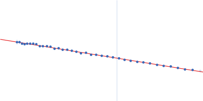
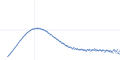
SAXS
data from solutions of
Plasminogen Roche
in
10mM Na2HPO4, 1.8 mM KH2PO4, 2.7 mM KCl, 137 mM NaCl, pH 7.4
were collected
on the
Rigaku BioSAXS-2000 instrument (CEITEC, Brno, Czech Republic)
using a Rigaku HyPix-3000 detector
at a sample-detector distance of 0.5 m and
at a wavelength of λ = 0.154 nm
(I(s) vs s, where s = 4πsinθ/λ, and 2θ is the scattering angle).
One solute concentration of 2.00 mg/ml was measured
at 20°C.
Six successive
600 second frames were collected.
The data were normalized to the intensity of the transmitted beam and radially averaged; the scattering of the solvent-blank was subtracted.

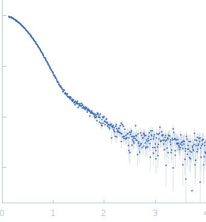 s, nm-1