| MWI(0) | 100 | kDa |
| MWexpected | 102 | kDa |
| VPorod | 157 | nm3 |
|
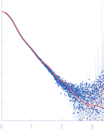
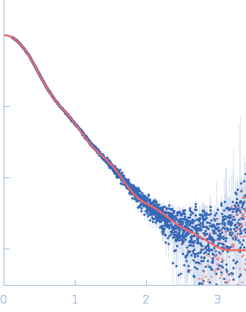
log I(s)
1.20×10⁻¹
1.20×10⁻²
1.20×10⁻³
1.20×10⁻⁴
|
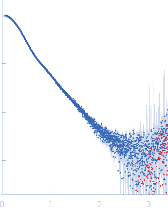 s, nm-1
|
|
|
|

|
|

|
|
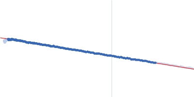
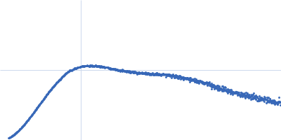
Synchrotron SAXS data were collected on the B21 beam line at the Diamond Light Source storage ring (Didcot, UK) using an Eiger 4M detector at a sample-detector distance of 3.7 m and at a wavelength of λ = 0.09524 nm (I(s) vs s, where s = 4πsinθ/λ, and 2θ is the scattering angle). Data were collected with an in-line size exclusion chromatography column (Shodex KW403, Shanghai, China) equilibrated in 20mM Sodium Phosphate, pH7.4, 150mM sodium chloride. 35μL of sample at a concentration of ~5 mg/ml was run at a flowrate of 0.16 ml/min and 3 s X-ray exposures were collected continuously during a 30min elution, resulting in 619 frames. Images were corrected, normalised and processed into 1D curves using GDA and DAWN at the Diamond Light Source.
|
|
|||||||||||||||||||||