Glutathione S -Transferase Mediated Epoxide Conversion: Functional and Structural Properties of an Enantioselective Catalyst

Haarmann M, Scholz M, Lienkamp A, Burnik J, Justesen B, Reiners J, Maier A, Eggerichs D, Vocke K, Smits S, Günther Pomorski T, Hofmann E, Tischler D, ACS Catalysis :12308-12324 (2025) DOI

SASDQY7 – StyI glutathione S-transferase

GST N-terminal domain-containing protein
MWI(0) 61 kDa
MWexpected 60 kDa
VPorod 91 nm3
log I(s) 4.43×10⁻² 4.43×10⁻³ 4.43×10⁻⁴ 4.43×10⁻⁵
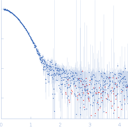
GST N-terminal domain-containing protein small angle scattering data  s, nm-1
ln I(s)
GST N-terminal domain-containing protein Guinier plot ln 4.44×10⁻² Rg: 2.7 nm 0 (2.7 nm)-2 s2
(sRg)2I(s)/I(0)
GST N-terminal domain-containing protein Kratky plot 1.104 0 3 sRg
p(r)
GST N-terminal domain-containing protein pair distance distribution function Rg: 2.7 nm 0 Dmax: 9.2 nm

Data validation


Fits and models


log I(s)
 s, nm-1
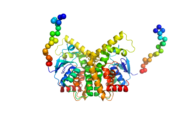
GST N-terminal domain-containing protein CORAL model

SAXS data from solutions of StyI glutathione S-transferase in 10 mM Tris, 300 mM NaCl, pH 8 were collected using a Xenocs Xeuss 2.0 Q-Zoom instrument equipped with a Pilatus3 R 300 K detector and a GENIX 3D CU Ultra Low Divergence X-ray beam delivery system (Center for Structural Studies, Heinrich-Heine-University, Düsseldorf, Germany) at a sample-detector distance of 0.6 m and at a wavelength of λ = 0.154 nm (I(s) vs s, where s = 4πsinθ/λ, and 2θ is the scattering angle). One solute concentration of 4.70 mg/ml was measured at 10°C. 12 successive 600 second frames were collected. The data were normalized to the intensity of the transmitted beam and radially averaged; the scattering of the solvent-blank was subtracted.

GST N-terminal domain-containing protein (Styl)
Mol. type   Protein
Organism   Gordonia rubripertincta
Olig. state   Dimer
Mon. MW   29.8 kDa
 
UniProt   A0A222TSG8 (1-238)
Sequence   FASTA