Dynamics and structural features of the eEF1A1 and eEF1A2 paralogs

Novosylna O, Shalak V, Dąbrowska K, Patmanidis I, Lozhko D, Bondarchuk T, Schiøtt B, Pedersen J, Knudsen C, Nissen P, Dadlez M, Negrutskii B, Nucleic Acids Research 53(21) (2025) DOI

SASDXH2 – Mammalian translation elongation factor eEF1A2

Mammalian translation elongation factor eEF1A2
MWI(0) 63 kDa
MWexpected 50 kDa
VPorod 60 nm3
log I(s) 7.98×10⁻² 7.98×10⁻³ 7.98×10⁻⁴ 7.98×10⁻⁵
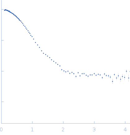
Mammalian translation elongation factor eEF1A2 small angle scattering data  s, nm-1
ln I(s)
Mammalian translation elongation factor eEF1A2 Guinier plot ln 7.99×10⁻² Rg: 2.6 nm 0 (2.6 nm)-2 s2
(sRg)2I(s)/I(0)
Mammalian translation elongation factor eEF1A2 Kratky plot 1.104 0 3 sRg
p(r)
Mammalian translation elongation factor eEF1A2 pair distance distribution function Rg: 26.7 nm 0 Dmax: 97.1 nm

Data validation


Fits and models


log I(s)
 s, nm-1
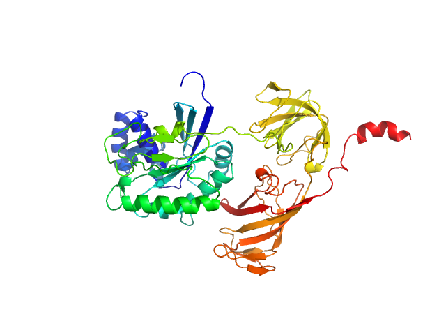
Mammalian translation elongation factor eEF1A2 CHIMERA model

SAXS data from solutions of Mammalian translation elongation factor eEF1A2 in 25 mM Tris HCl, 150 mM NaCl, 6 mM βME, 20% glycerol, 0.01mM GDP,, pH 7.5 were collected on the Bruker Nanostar w Excillum source instrument (Department of Chemistry, iNANO building, Aarhus Uinversity, Aarhus C, Denmark) using a VÅNTEC-2000 detector at a sample-detector distance of 0.9 m and at a wavelength of λ = 0.134 nm (I(s) vs s, where s = 4πsinθ/λ, and 2θ is the scattering angle). One solute concentration of 1.90 mg/ml was measured at 20°C. One 1800 second frame was collected. The data were normalized to the intensity of the transmitted beam and radially averaged; the scattering of the solvent-blank was subtracted.

Mammalian translation elongation factor eEF1A2
Mol. type   Protein
Olig. state   Monomer
Mon. MW   50.5 kDa
 
UniProt   Q05639 (1-463)
Sequence   FASTA