Open or closed? Structural characterization of plasma-derived and recombinant human plasminogen across multiple resolution scales.

Kasiarova L, Verma N, Pinkas M, Henek T, Klumpler T Chovancikova M, Reutova H, Kasparek P, Hlozankova M, Damborsky J, Novacek J, Hernychova L, Marek M, Int J Biol Macromol :154885 (2026) Europe PMC

SASDZ24 – Plasminogen R561A point mutant

Plasminogen R561A - pointmutant
MWexperimental 79 kDa
MWexpected 88 kDa
VPorod 162 nm3
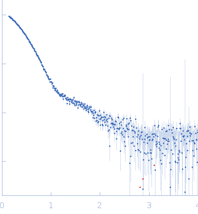
log I(s) 1.84×100 1.84×10⁻¹ 1.84×10⁻² 1.84×10⁻³
Plasminogen R561A - pointmutant small angle scattering data  s, nm-1
ln I(s)
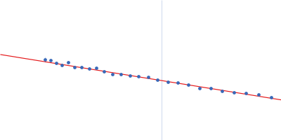
Plasminogen R561A - pointmutant Guinier plot ln 1.84×100 Rg: 3.7 nm 0 (3.7 nm)-2 s2
(sRg)2I(s)/I(0)
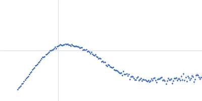
Plasminogen R561A - pointmutant Kratky plot 1.104 0 √3 sRg
p(r)
Plasminogen R561A - pointmutant pair distance distribution function Rg: 3.8 nm 0 Dmax: 12.4 nm

Data validation


There are no models related to this curve.

SAXS data from solutions of Plasminogen R561A point mutant in 10mM Na2HPO4, 1.8 mM KH2PO4, 2.7 mM KCl, 137 mM NaCl, pH 7.4 were collected on the Rigaku BioSAXS-2000 instrument (CEITEC, Brno, Czech Republic) using a Rigaku HyPix-3000 detector at a sample-detector distance of 0.5 m and at a wavelength of λ = 0.154 nm (I(s) vs s, where s = 4πsinθ/λ, and 2θ is the scattering angle). One solute concentration of 3.00 mg/ml was measured at 20°C. Six successive 600 second frames were collected. The data were normalized to the intensity of the transmitted beam and radially averaged; the scattering of the solvent-blank was subtracted.

Plasminogen R561A - pointmutant (PLG R561A)
Mol. type   Protein
Organism   Homo sapiens, production HEK298 EBNA cell line
Olig. state   Monomer
Mon. MW   88.3 kDa
 
UniProt   P00747 (20-810)
Sequence   FASTA