VirB8 homolog TraE from plasmid pKM101 forms a hexameric ring structure and interacts with the VirB6 homolog TraD.

Casu B, Mary C, Sverzhinsky A, Fouillen A, Nanci A, Baron C, Proc Natl Acad Sci U S A 115(23):5950-5955 (2018) Europe PMC

SASDB75 – Escherichia coli TraE protein: A VirB8 homolog from plasmid pKM101

Escherichia coli TraE protein (VirB8 homolog)
MWexperimental 264 kDa
MWexpected 171 kDa
VPorod 360 nm3
log I(s) 9.40×10⁻³ 9.40×10⁻⁴ 9.40×10⁻⁵ 9.40×10⁻⁶
Escherichia coli TraE protein (VirB8 homolog) small angle scattering data  s, nm-1
ln I(s)
Escherichia coli TraE protein (VirB8 homolog) Guinier plot ln 9.40×10⁻³ Rg: 4.4 nm 0 (4.4 nm)-2 s2
(sRg)2I(s)/I(0)
Escherichia coli TraE protein (VirB8 homolog) Kratky plot 1.104 0 3 sRg
p(r)
Escherichia coli TraE protein (VirB8 homolog) pair distance distribution function Rg: 4.4 nm 0 Dmax: 13.7 nm

Data validation


Fits and models


log I(s)
 s, nm-1
Escherichia coli TraE protein (VirB8 homolog) GASBOR model

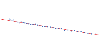
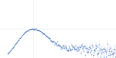
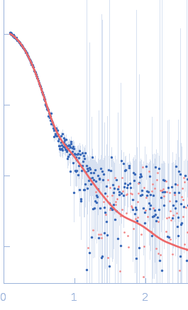
X-ray synchrotron radiation scattering data from a solution of E. coli TraE protein in 50 mM sodium phosphate, 300 mM NaCl, 40 mM imidazole, 0.15 % octyl glucose neopentyl glycol (OGNG), pH 7.4 were collected using size-exclusion chromatography (SEC) SAXS on the G1 beam line at the CHESS storage ring (Ithaca, USA) using a CCD Finger Lakes CCD detector (I(s) vs s, where s = 4π sin θ/λ; 2θ is the scattering angle; λ = 0.12 nm). The data were collected as 70 successive 2 second frames through the hexameric TraE protein/OGNG elution peak and were normalized to the intensity of the transmitted beam and radially averaged. The scattering of the solvent-blank was subtracted to produce the scattering profile displayed in this entry.

The MW of the TraE/OGNG complex was additionally confirmed using SEC-MALLS (248kDa).
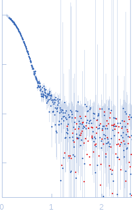
Escherichia coli TraE protein (VirB8 homolog) (TraE)
Mol. type   Protein
Organism   Escherichia coli
Olig. state   Hexamer
Mon. MW   28.5 kDa
 
UniProt   Q46703
Sequence   FASTA