Direct interaction of DNA repair protein tyrosyl DNA phosphodiesterase 1 and the DNA ligase III catalytic domain is regulated by phosphorylation of its flexible N-terminus.

Rashid I, Hammel M, Sverzhinsky A, Tsai MS, Pascal JM, Tainer JA, Tomkinson AE, J Biol Chem :100921 (2021) Europe PMC

SASDJS2 – Truncated DNA ligase 3 bound to tyrosyl-DNA phosphodiesterase 1 (LigIIIα(170-755)/TDP1 complex)

Tyrosyl-DNA phosphodiesterase 1
Isoform 3 of DNA ligase 3 (DNA ligase III alpha)
MWexperimental 140 kDa
MWexpected 140 kDa
VPorod 211 nm3
log I(s) 7.49×101 7.49×100 7.49×10⁻¹ 7.49×10⁻²
Tyrosyl-DNA phosphodiesterase 1 Isoform 3 of DNA ligase 3 (DNA ligase III alpha) small angle scattering data  s, nm-1
ln I(s)
Tyrosyl-DNA phosphodiesterase 1 Isoform 3 of DNA ligase 3 (DNA ligase III alpha) Guinier plot ln 7.50×101 Rg: 4.5 nm 0 (4.5 nm)-2 s2
(sRg)2I(s)/I(0)
Tyrosyl-DNA phosphodiesterase 1 Isoform 3 of DNA ligase 3 (DNA ligase III alpha) Kratky plot 1.104 0 3 sRg
p(r)
Tyrosyl-DNA phosphodiesterase 1 Isoform 3 of DNA ligase 3 (DNA ligase III alpha) pair distance distribution function Rg: 4.9 nm 0 Dmax: 18 nm

Data validation


Fits and models


log I(s)
 s, nm-1
Tyrosyl-DNA phosphodiesterase 1 Isoform 3 of DNA ligase 3 (DNA ligase III alpha) BILBOMD model

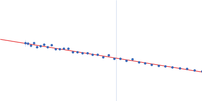
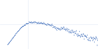
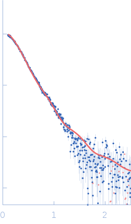
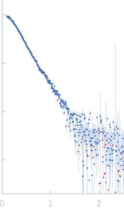
Synchrotron SAXS data from solutions of the LigIIIα(170-755)/TDP1 complex in 200 mM NaCl, 40 mM HEPES, pH 7.5 were collected on the 12.3.1 (SIBYLS) beam line at the Advanced Light Source (ALS; Berkeley, CA, USA) using a Pilatus3 X 2M detector at a sample-detector distance of 2.1 m and at a wavelength of λ = 0.1127 nm (I(s) vs s, where s = 4πsinθ/λ, and 2θ is the scattering angle). In-line size-exclusion chromatography (SEC) SAS was employed. The SEC parameters were as follows: A 50.00 μl sample at 3 mg/ml was injected at a 0.50 ml/min flow rate onto a Shodex KW-800 series column at 20°C. 600 successive 3 second frames were collected. The data were normalized to the intensity of the transmitted beam and radially averaged; the scattering of the solvent-blank was subtracted.

Tyrosyl-DNA phosphodiesterase 1 (TDP1)
Mol. type   Protein
Organism   Homo sapiens
Olig. state   Monomer
Mon. MW   71.2 kDa
 
UniProt   Q9NUW8 (1-608)
Sequence   FASTA
 
Isoform 3 of DNA ligase 3 (DNA ligase III alpha) (LigIIIα(170-755))
Mol. type   Protein
Organism   Homo sapiens
Olig. state   Monomer
Mon. MW   68.3 kDa
 
UniProt   P49916-3 (170-755)
Sequence   FASTA