Integrated Structural Modeling of Full-Length LRH-1 Reveals Inter-domain Interactions Contribute to Receptor Structure and Function.

Seacrist CD, Kuenze G, Hoffmann RM, Moeller BE, Burke JE, Meiler J, Blind RD, Structure (2020) Europe PMC

SASDG85 – Wild-type full-length Liver Receptor Homolog-1/Peroxisome Proliferator-Activated Receptor Gamma Coactivator-1 Alpha peptide complex bound to the CYP7A1 promoter oligonucleotide duplex.

Liver Receptor Homolog-1
Peroxisome Proliferator-Activated Receptor Gamma Coactivator-1 Alpha
CYP7A1 Promoter Forward
CYP7A1 Promoter Reverse
MWexperimental 67 kDa
MWexpected 73 kDa
VPorod 76 nm3
log I(s) 2.38×101 2.38×100 2.38×10⁻¹ 2.38×10⁻²
Liver Receptor Homolog-1 Peroxisome Proliferator-Activated Receptor Gamma Coactivator-1 Alpha CYP7A1 Promoter Forward CYP7A1 Promoter Reverse small angle scattering data  s, nm-1
ln I(s)
Liver Receptor Homolog-1 Peroxisome Proliferator-Activated Receptor Gamma Coactivator-1 Alpha CYP7A1 Promoter Forward CYP7A1 Promoter Reverse Guinier plot ln 2.39×101 Rg: 3.8 nm 0 (3.8 nm)-2 s2
(sRg)2I(s)/I(0)
Liver Receptor Homolog-1 Peroxisome Proliferator-Activated Receptor Gamma Coactivator-1 Alpha CYP7A1 Promoter Forward CYP7A1 Promoter Reverse Kratky plot 1.104 0 3 sRg
p(r)
Liver Receptor Homolog-1 Peroxisome Proliferator-Activated Receptor Gamma Coactivator-1 Alpha CYP7A1 Promoter Forward CYP7A1 Promoter Reverse pair distance distribution function Rg: 3.9 nm 0 Dmax: 13 nm

Data validation


There are no models related to this curve.

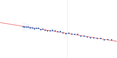
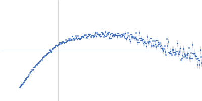
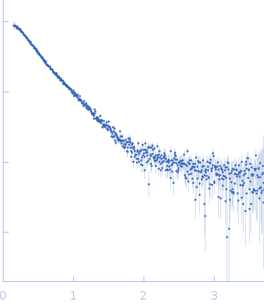
Synchrotron SAXS data from solutions of WT flLRH-1 / CYP7A1 oligo duplex / PGC1 alpha co-activator peptide in 20 mM TRIS, 150 mM NaCl, 2% v/v glycerol, 0.5 mM CHAPS, 5 mM DTT, pH 7.5 were collected on the SIBYLS 12.3.1 beam line at the Advanced Light Source (ALS; Berkeley, CA, USA) using a Pilatus3 X 2M detector at a sample-detector distance of 2.1 m and at a wavelength of λ = 0.1127 nm (I(s) vs s, where s = 4πsinθ/λ, and 2θ is the scattering angle). In-line size-exclusion chromatography (SEC) SAS was employed. The SEC parameters were as follows: A 100.00 μl sample at 7.5 mg/ml was injected at a 0.45 ml/min flow rate onto a Shodex KW-800 series column at 20°C. 500 successive 3 second frames were collected continuously during the ~25 minute elution. After 2D-to-1D radial averaging, the data were analyzed through the SEC elution peak of the sample (from which I(0) and Rg were evaluated using the Guinier approximation) and the scattering of an appropriate solvent-blank was subtracted.

Storage temperature = UNKNOWN

Liver Receptor Homolog-1 (LRH-1)
Mol. type   Protein
Organism   Homo sapiens
Olig. state   Monomer
Mon. MW   64.1 kDa
 
UniProt   O00482 (1-541)
Sequence   FASTA
 
Peroxisome Proliferator-Activated Receptor Gamma Coactivator-1 Alpha (PGC1 alpha)
Mol. type   Protein
Organism   Homo sapiens
Olig. state   Monomer
Mon. MW   1.5 kDa
Sequence   FASTA
 
CYP7A1 Promoter Forward
Mol. type   DNA
Organism   Homo sapiens
Olig. state   Monomer
Mon. MW   3.8 kDa
Sequence   FASTA
 
CYP7A1 Promoter Reverse
Mol. type   DNA
Organism   Homo sapiens
Olig. state   Monomer
Mon. MW   3.7 kDa
Sequence   FASTA