| MWexperimental | 52 | kDa |
| MWexpected | 51 | kDa |
| VPorod | 74 | nm3 |
|
log I(s)
7.35×10⁻²
7.35×10⁻³
7.35×10⁻⁴
7.35×10⁻⁵
|
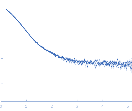 s, nm-1
|
|
|
|

|
|

|
|
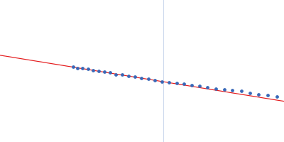
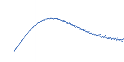
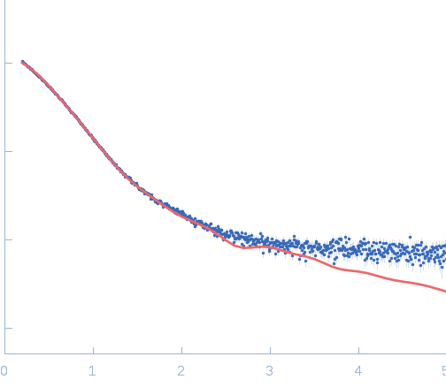
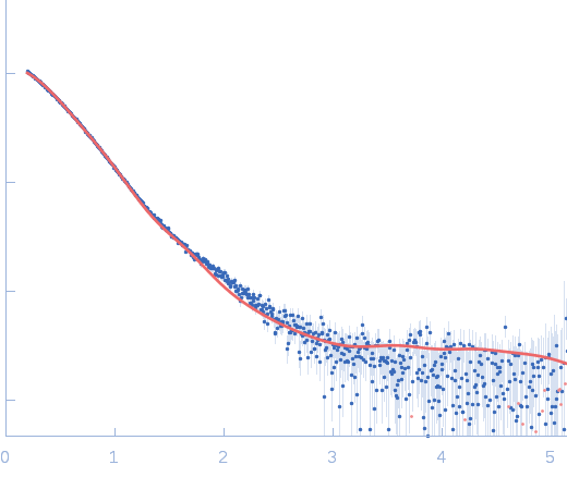
Synchrotron SAXS data from solutions of Vaccinia virus MVA F1L antiapoptotic Bcl-2 viral protein in 25 mM HEPES, 150 mM NaCl, 5 mM DTT, pH 7.5 were collected on the SAXS/WAXS camera on the storage ring Australian Synchrotron (Melbourne, Australia) using a Pilatus 1M detector at a sample-detector distance of 1.6 m (I(s) vs s = 4π sin θ/λ, where 2θ is the scattering angle). Solute concentrations ranging between 0.1 and 3.6 mg/ml were measured. 18 successive 1 second frames were collected. The data were normalized to the intensity of the transmitted beam and radially averaged; the scattering of the solvent-blank was subtracted. The low angle data collected were obtained from a single concentration scattering curve.
Wavelength = UNKNOWN. Cell temperature = UNKNOWN |
|
|||||||||||||||||||||||||||