Solution structure of the type I polyketide synthase Pks13 from Mycobacterium tuberculosis.

Bon C, Cabantous S, Julien S, Guillet V, Chalut C, Rima J, Brison Y, Malaga W, Sanchez-Dafun A, Gavalda S, Quémard A, Marcoux J, Waldo GS, Guilhot C, Mourey L, BMC Biol 20(1):147 (2022) Europe PMC

SASDNU9 – Mycocerosic acid synthase in its apo form and at neutral pH

Mycocerosic acid synthase
MWI(0) 221 kDa
MWexpected 224 kDa
VPorod 420 nm3
log I(s) 1.27×10⁻¹ 1.27×10⁻² 1.27×10⁻³ 1.27×10⁻⁴
Mycocerosic acid synthase small angle scattering data  s, nm-1
ln I(s)
Mycocerosic acid synthase Guinier plot ln 1.28×10⁻¹ Rg: 5.7 nm 0 (5.7 nm)-2 s2
(sRg)2I(s)/I(0)
Mycocerosic acid synthase Kratky plot 1.104 0 3 sRg
p(r)
Mycocerosic acid synthase pair distance distribution function Rg: 5.9 nm 0 Dmax: 21 nm

Data validation


Fits and models


log I(s)
 s, nm-1
Mycocerosic acid synthase DAMMIN model

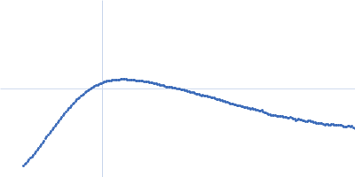
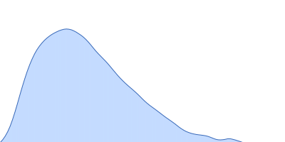
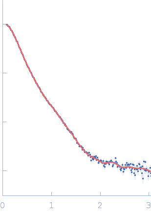
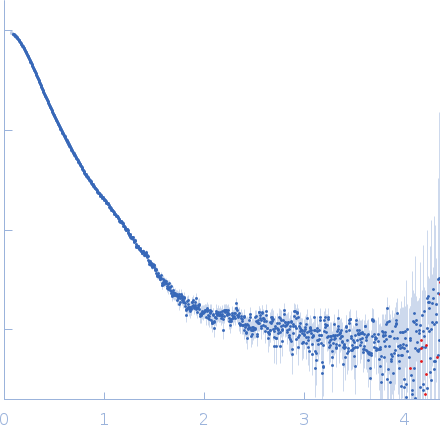
Synchrotron SAXS data from solutions of mycocerosic acid synthase in its apo form and at neutral pH in 50 mM Tris-HCl, 50 mM NaCl, 10% glycerol, pH 8 were collected on the SWING beam line at the SOLEIL storage ring (Saint-Aubin, France) using a AVIEX PCCD170170 detector at a sample-detector distance of 2.5 m and at a wavelength of λ = 0.1033 nm (I(s) vs s, where s = 4πsinθ/λ, and 2θ is the scattering angle). In-line size-exclusion chromatography (SEC) SAS was employed. The SEC parameters were as follows: A 40.00 μl sample at 3 mg/ml was injected at a 0.15 ml/min flow rate onto a Agilent Bio SEC-3, 300 Å column at 12°C. The data were normalized to the intensity of the transmitted beam and radially averaged; the scattering of the solvent-blank was subtracted.

Number of frames = UNKNOWN

Mycocerosic acid synthase (MAS)
Mol. type   Protein
Organism   Mycobacterium bovis (strain ATCC BAA-935 / AF2122/97)
Olig. state   Monomer
Mon. MW   224.4 kDa
 
UniProt   Q02251 (1-2111)
Sequence   FASTA