|
|
|
|
|
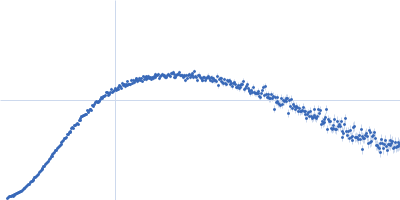
| Sample: |
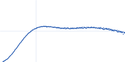
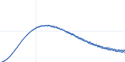
Heat shock cognate 71 kDa protein, 71 kDa protein
|
| Buffer: |
20 mM HEPES, 150 mM KCl, 10 mM M gCl2, 1mM TCEP, pH: 7.5 |
| Experiment: |
SAXS
data collected at EMBL P12, PETRA III on 2025 Mar 6
|
Advances on the large-scale bacterial production and stable phosphorylation of Hsc70
Protein Expression and Purification :106923 (2026)
Kley N, Hecht-Bucher M, Itzen A
|
| RgGuinier |
5.7 |
nm |
| Dmax |
24.5 |
nm |
| VolumePorod |
351 |
nm3 |
|
|
|
|
|
|
|
| Sample: |
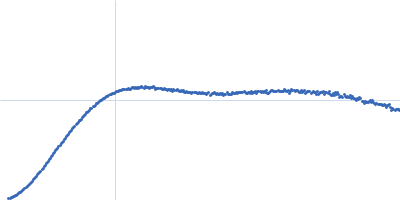
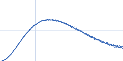
Heat shock cognate 71 kDa protein, 71 kDa protein
|
| Buffer: |
20 mM HEPES, 150 mM KCl, 10 mM M gCl2, 1mM TCEP, pH: 7.5 |
| Experiment: |
SAXS
data collected at EMBL P12, PETRA III on 2025 Mar 6
|
Advances on the large-scale bacterial production and stable phosphorylation of Hsc70
Protein Expression and Purification :106923 (2026)
Kley N, Hecht-Bucher M, Itzen A
|
| RgGuinier |
5.7 |
nm |
| Dmax |
24.7 |
nm |
| VolumePorod |
352 |
nm3 |
|
|
|
|
|
|
|
| Sample: |
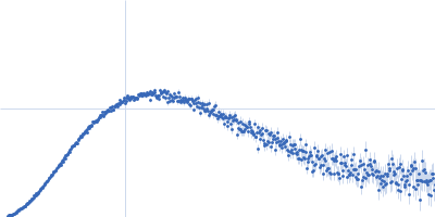
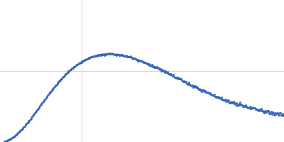
Heat shock cognate 71 kDa protein, 71 kDa protein
|
| Buffer: |
20 mM HEPES, 150 mM KCl, 10 mM M gCl2, 1mM TCEP, pH: 7.5 |
| Experiment: |
SAXS
data collected at EMBL P12, PETRA III on 2025 Mar 6
|
Advances on the large-scale bacterial production and stable phosphorylation of Hsc70
Protein Expression and Purification :106923 (2026)
Kley N, Hecht-Bucher M, Itzen A
|
| RgGuinier |
5.6 |
nm |
| Dmax |
25.0 |
nm |
| VolumePorod |
360 |
nm3 |
|
|
|
|
|
|
|
| Sample: |
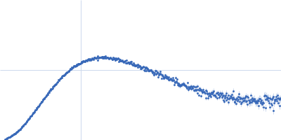
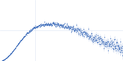
Phosphorylated Heat shock cognate 71 kDa protein, 71 kDa Homo sapiens protein
|
| Buffer: |
20 mM HEPES, 150 mM KCl, 10 mM MgCl2, 1 mM TCEP, 1 mM ATP, pH: 7.5 |
| Experiment: |
SAXS
data collected at EMBL P12, PETRA III on 2025 Mar 6
|
Advances on the large-scale bacterial production and stable phosphorylation of Hsc70
Protein Expression and Purification :106923 (2026)
Kley N, Hecht-Bucher M, Itzen A
|
| RgGuinier |
3.6 |
nm |
| Dmax |
15.3 |
nm |
| VolumePorod |
122 |
nm3 |
|
|
|
|
|
|
|
| Sample: |
Phosphorylated Heat shock cognate 71 kDa protein, 71 kDa Homo sapiens protein
|
| Buffer: |
20 mM HEPES, 150 mM KCl, 10 mM MgCl2, 1 mM TCEP, 1 mM ATP, pH: 7.5 |
| Experiment: |
SAXS
data collected at EMBL P12, PETRA III on 2025 Mar 6
|
Advances on the large-scale bacterial production and stable phosphorylation of Hsc70
Protein Expression and Purification :106923 (2026)
Kley N, Hecht-Bucher M, Itzen A
|
| RgGuinier |
3.7 |
nm |
| Dmax |
16.4 |
nm |
| VolumePorod |
131 |
nm3 |
|
|
|
|
|
|
|
| Sample: |
Phosphorylated Heat shock cognate 71 kDa protein, 71 kDa Homo sapiens protein
|
| Buffer: |
20 mM HEPES, 150 mM KCl, 10 mM MgCl2, 1 mM TCEP, 1 mM ATP, pH: 7.5 |
| Experiment: |
SAXS
data collected at EMBL P12, PETRA III on 2025 Mar 6
|
Advances on the large-scale bacterial production and stable phosphorylation of Hsc70
Protein Expression and Purification :106923 (2026)
Kley N, Hecht-Bucher M, Itzen A
|
| RgGuinier |
3.9 |
nm |
| Dmax |
16.7 |
nm |
| VolumePorod |
141 |
nm3 |
|
|
|
|
|
|
|
| Sample: |
Phosphorylated Heat shock cognate 71 kDa protein, 71 kDa Homo sapiens protein
|
| Buffer: |
20 mM HEPES, 150 mM KCl, 10 mM MgCl2, 1 mM TCEP, 1 mM ATP, pH: 7.5 |
| Experiment: |
SAXS
data collected at EMBL P12, PETRA III on 2025 Mar 6
|
Advances on the large-scale bacterial production and stable phosphorylation of Hsc70
Protein Expression and Purification :106923 (2026)
Kley N, Hecht-Bucher M, Itzen A
|
| RgGuinier |
4.3 |
nm |
| Dmax |
16.7 |
nm |
| VolumePorod |
149 |
nm3 |
|
|
|
|
|
|
|
| Sample: |
Phosphorylated Heat shock cognate 71 kDa protein, 71 kDa Homo sapiens protein
|
| Buffer: |
20 mM HEPES, 150 mM KCl, 10 mM MgCl2, 1 mM TCEP, 1 mM ATP, pH: 7.5 |
| Experiment: |
SAXS
data collected at EMBL P12, PETRA III on 2025 Mar 6
|
Advances on the large-scale bacterial production and stable phosphorylation of Hsc70
Protein Expression and Purification :106923 (2026)
Kley N, Hecht-Bucher M, Itzen A
|
| RgGuinier |
4.2 |
nm |
| Dmax |
18.1 |
nm |
| VolumePorod |
162 |
nm3 |
|
|
|
|
|
|
|
| Sample: |
Phosphorylated Heat shock cognate 71 kDa protein, 71 kDa Homo sapiens protein
|
| Buffer: |
20 mM HEPES, 150 mM KCl, 10 mM MgCl2, 1mM TCEP, 1mM ADP, pH: 7.5 |
| Experiment: |
SAXS
data collected at EMBL P12, PETRA III on 2025 Mar 6
|
Advances on the large-scale bacterial production and stable phosphorylation of Hsc70
Protein Expression and Purification :106923 (2026)
Kley N, Hecht-Bucher M, Itzen A
|
| RgGuinier |
4.4 |
nm |
| Dmax |
20.3 |
nm |
| VolumePorod |
169 |
nm3 |
|
|
|
|
|
|
|
| Sample: |
Phosphorylated Heat shock cognate 71 kDa protein, 71 kDa Homo sapiens protein
|
| Buffer: |
20 mM HEPES, 150 mM KCl, 10 mM MgCl2, 1mM TCEP, 1mM ADP, pH: 7.5 |
| Experiment: |
SAXS
data collected at EMBL P12, PETRA III on 2025 Mar 6
|
Advances on the large-scale bacterial production and stable phosphorylation of Hsc70
Protein Expression and Purification :106923 (2026)
Kley N, Hecht-Bucher M, Itzen A
|
| RgGuinier |
4.8 |
nm |
| Dmax |
22.3 |
nm |
| VolumePorod |
203 |
nm3 |
|
|