|
|
|
|
|
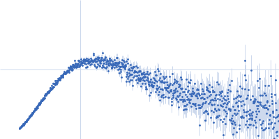
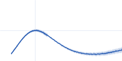
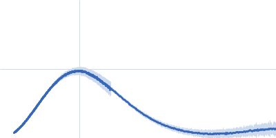
| Sample: |
Ferric anguibactin-binding protein monomer, 32 kDa Bacillus cereus (strain … protein
|
| Buffer: |
20mM Tris-HCl, 100 mM NaCl, pH: 8 |
| Experiment: |
SAXS
data collected at 4C, Pohang Accelerator Laboratory on 2025 Apr 7
|
FatB
|
| RgGuinier |
2.2 |
nm |
| Dmax |
7.4 |
nm |
| VolumePorod |
66 |
nm3 |
|
|
|
|
|
|
|
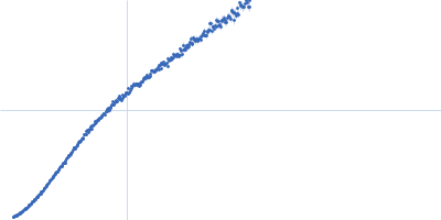
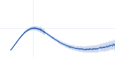
| Sample: |
Ferric anguibactin-binding protein monomer, 32 kDa Bacillus cereus (strain … protein
|
| Buffer: |
20mM Tris-HCl, 100 mM NaCl, pH: 8 |
| Experiment: |
SAXS
data collected at 4C, Pohang Accelerator Laboratory on 2025 Apr 7
|
FatB
|
| RgGuinier |
2.2 |
nm |
| Dmax |
7.5 |
nm |
| VolumePorod |
65 |
nm3 |
|
|
|
|
|
|
|
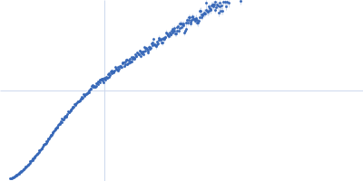
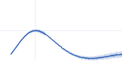
| Sample: |
Ferric anguibactin-binding protein monomer, 32 kDa Bacillus cereus (strain … protein
|
| Buffer: |
20mM Tris-HCl, 100 mM NaCl, pH: 8 |
| Experiment: |
SAXS
data collected at 4C, Pohang Accelerator Laboratory on 2025 Apr 7
|
FatB
|
| RgGuinier |
2.1 |
nm |
| Dmax |
7.1 |
nm |
| VolumePorod |
56 |
nm3 |
|
|
|
|
|
|
|
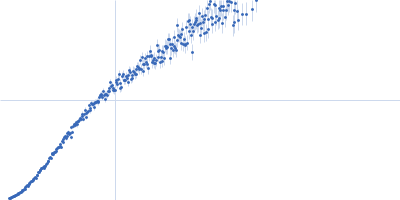
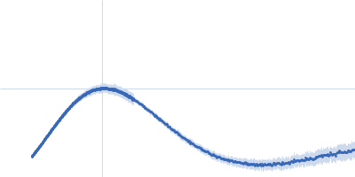
| Sample: |
Ferric anguibactin-binding protein monomer, 32 kDa Bacillus cereus (strain … protein
|
| Buffer: |
20mM Tris-HCl, 100 mM NaCl, pH: 8 |
| Experiment: |
SAXS
data collected at 4C, Pohang Accelerator Laboratory on 2025 Apr 7
|
FatB
|
| RgGuinier |
2.1 |
nm |
| Dmax |
7.1 |
nm |
| VolumePorod |
57 |
nm3 |
|
|
|
|
|
|
|
| Sample: |
Ferric anguibactin-binding protein monomer, 32 kDa Bacillus cereus (strain … protein
|
| Buffer: |
20mM Tris-HCl, 100 mM NaCl, pH: 8 |
| Experiment: |
SAXS
data collected at 4C, Pohang Accelerator Laboratory on 2025 Apr 7
|
FatB
|
| RgGuinier |
2.1 |
nm |
| Dmax |
7.1 |
nm |
| VolumePorod |
42 |
nm3 |
|
|
|
|
|
|
|
| Sample: |
Mutated ribosome assembly protein 1 (R1086Q) with C-terminal tag (RSRSGSENLYFQGSHHHHHHHH) monomer, 127 kDa Saccharomyces cerevisiae protein
|
| Buffer: |
50 mM HEPES, 300 mM NaCl, 5 mM MgCl2, 5% glycerol, pH: 8 |
| Experiment: |
SAXS
data collected at B21, Diamond Light Source on 2024 Oct 3
|
Hydroxyl radical footprinting modification reveals an intradomain communication pathway in EFL1 disrupted by a Shwachman-Diamond syndrome-associated mutation.
Protein Sci 35(4):e70504 (2026)
Zúñiga-Domínguez JA, Jain R, González-Andrade M, Farquhar ER, Chance MR, Gijsbers A, Sánchez-Puig N
|
| RgGuinier |
4.7 |
nm |
| Dmax |
15.2 |
nm |
| VolumePorod |
314 |
nm3 |
|
|
|
|
|
|
|
| Sample: |
Ribosome assembly protein 1 with C-terminal tag (RSRSGSENLYFQGSHHHHHHHH) monomer, 127 kDa Saccharomyces cerevisiae protein
|
| Buffer: |
50 mM HEPES, 300 mM NaCl, 5 mM MgCl2, 5% glycerol, pH: 8 |
| Experiment: |
SAXS
data collected at B21, Diamond Light Source on 2024 Oct 3
|
Hydroxyl radical footprinting modification reveals an intradomain communication pathway in EFL1 disrupted by a Shwachman-Diamond syndrome-associated mutation.
Protein Sci 35(4):e70504 (2026)
Zúñiga-Domínguez JA, Jain R, González-Andrade M, Farquhar ER, Chance MR, Gijsbers A, Sánchez-Puig N
|
| RgGuinier |
4.6 |
nm |
| Dmax |
15.7 |
nm |
| VolumePorod |
368 |
nm3 |
|
|
|
|
|
|
|
| Sample: |
Cellular tumor antigen p53 monomer, 4 kDa Homo sapiens protein
|
| Buffer: |
20 mM Tris, 150 mM NaCl,, pH: 7 |
| Experiment: |
SAXS
data collected at BM29, ESRF on 2025 Jan 30
|
Conformational Flexibility and Transient Structure of the Proline-Rich Domain in p53.
Biophys J (2026)
Berggren A, Bakker M, Fisher H, Skepö M
|
|
|
|
|
|
|
|
| Sample: |
Cellular tumor antigen p53 monomer, 4 kDa Homo sapiens protein
|
| Buffer: |
20 mM Tris, 10 mM NaCl,, pH: 7 |
| Experiment: |
SAXS
data collected at BM29, ESRF on 2025 Jan 30
|
Conformational Flexibility and Transient Structure of the Proline-Rich Domain in p53.
Biophys J (2026)
Berggren A, Bakker M, Fisher H, Skepö M
|
|
|
|
|
|
|
|
| Sample: |
Cellular tumor antigen p53 P72R monomer, 4 kDa Homo sapiens protein
|
| Buffer: |
20 mM Tris, 150 mM NaCl,, pH: 7 |
| Experiment: |
SAXS
data collected at BM29, ESRF on 2025 Sep 25
|
Conformational Flexibility and Transient Structure of the Proline-Rich Domain in p53.
Biophys J (2026)
Berggren A, Bakker M, Fisher H, Skepö M
|
|
|


 with C-terminal tag (RSRSGSENLYFQGSHHHHHHHH) experimental SAS data")

 experimental SAS data")