|
|
|
|
|
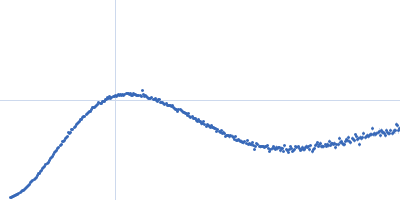
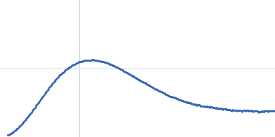
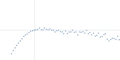
| Sample: |
Type II secretion system protein L, periplasmic domain dimer, 28 kDa Pseudomonas aeruginosa protein
|
| Buffer: |
50 mM TRIS, 100 mM NaCl, pH: 7.5 |
| Experiment: |
SAXS
data collected at SWING, SOLEIL on 2016 Apr 8
|
Structure and oligomerization of the periplasmic domain of GspL from the type II secretion system of Pseudomonas aeruginosa.
Sci Rep 8(1):16760 (2018)
Fulara A, Vandenberghe I, Read RJ, Devreese B, Savvides SN
|
| RgGuinier |
3.2 |
nm |
| Dmax |
10.5 |
nm |
|
|
|
|
|
|
|
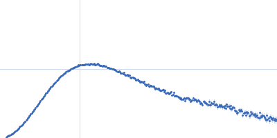
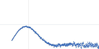
| Sample: |
Membrane scaffold protein 1D1 (deuterated, 75%) dimer, 49 kDa protein
1-palmitoyl-2-palmitoleoyl-sn-glycero-3-phosphocholine (deuteration: 78% head, 92% acyl), 1 kDa Escherichia coli
Calcium-transporting ATPase 8, plasma membrane-type monomer, 118 kDa Arabidopsis thaliana protein
|
| Buffer: |
30 mM Tris, 150 mM NaCl, 1mM MgCl2, 1 mM CaCl2, pH: 7.5 |
| Experiment: |
SANS
data collected at SANS-1, Heinz Maier-Leibnitz Zentrum on 2017 Aug 28
|
Structural basis for activation of plasma-membrane Ca2+-ATPase by calmodulin.
Commun Biol 1:206 (2018)
Nitsche J, Josts I, Heidemann J, Mertens HD, Maric S, Moulin M, Haertlein M, Busch S, Forsyth VT, Svergun DI, Uetrecht C, Tidow H
|
| RgGuinier |
4.0 |
nm |
| Dmax |
13.0 |
nm |
| VolumePorod |
202 |
nm3 |
|
|
|
|
|
|
|
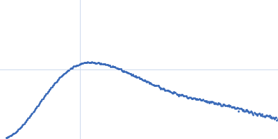
| Sample: |
Membrane scaffold protein 1D1 (deuterated, 75%) dimer, 49 kDa protein
1-palmitoyl-2-palmitoleoyl-sn-glycero-3-phosphocholine (deuteration: 78% head, 92% acyl), 1 kDa Escherichia coli
Calcium-transporting ATPase 8, plasma membrane-type monomer, 118 kDa Arabidopsis thaliana protein
Calmodulin-7 monomer, Arabidopsis thaliana protein
|
| Buffer: |
30 mM Tris, 150 mM NaCl, 1mM MgCl2, 1 mM CaCl2, pH: 7.5 |
| Experiment: |
SANS
data collected at SANS-1, Heinz Maier-Leibnitz Zentrum on 2017 Aug 28
|
Structural basis for activation of plasma-membrane Ca2+-ATPase by calmodulin.
Commun Biol 1:206 (2018)
Nitsche J, Josts I, Heidemann J, Mertens HD, Maric S, Moulin M, Haertlein M, Busch S, Forsyth VT, Svergun DI, Uetrecht C, Tidow H
|
| RgGuinier |
4.8 |
nm |
| Dmax |
18.0 |
nm |
| VolumePorod |
297 |
nm3 |
|
|
|
|
|
|
|
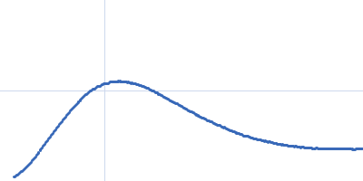
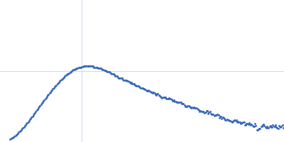
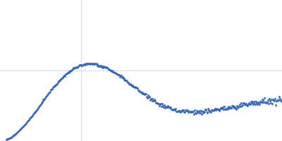
| Sample: |
Membrane scaffold protein 1D1 (deuterated, 75%) dimer, 49 kDa protein
1-palmitoyl-2-palmitoleoyl-sn-glycero-3-phosphocholine (deuteration: 78% head, 92% acyl), 1 kDa Escherichia coli
Calcium-transporting ATPase 8, plasma membrane-type monomer, 118 kDa Arabidopsis thaliana protein
|
| Buffer: |
30 mM Tris, 150 mM NaCl, 1mM MgCl2, 1 mM CaCl2, pH: 7.5 |
| Experiment: |
SAXS
data collected at EMBL P12, PETRA III on 2017 Sep 8
|
Structural basis for activation of plasma-membrane Ca2+-ATPase by calmodulin.
Commun Biol 1:206 (2018)
Nitsche J, Josts I, Heidemann J, Mertens HD, Maric S, Moulin M, Haertlein M, Busch S, Forsyth VT, Svergun DI, Uetrecht C, Tidow H
|
| RgGuinier |
5.3 |
nm |
| Dmax |
20.0 |
nm |
| VolumePorod |
626 |
nm3 |
|
|
|
|
|
|
|
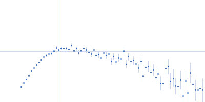
| Sample: |
Membrane scaffold protein 1D1 (deuterated, 75%) dimer, 49 kDa protein
1-palmitoyl-2-palmitoleoyl-sn-glycero-3-phosphocholine (deuteration: 78% head, 92% acyl), 1 kDa Escherichia coli
Calcium-transporting ATPase 8, plasma membrane-type monomer, 118 kDa Arabidopsis thaliana protein
Calmodulin-7 monomer, Arabidopsis thaliana protein
|
| Buffer: |
30 mM Tris, 150 mM NaCl, 1mM MgCl2, 1 mM CaCl2, pH: 7.5 |
| Experiment: |
SAXS
data collected at EMBL P12, PETRA III on 2017 Sep 8
|
Structural basis for activation of plasma-membrane Ca2+-ATPase by calmodulin.
Commun Biol 1:206 (2018)
Nitsche J, Josts I, Heidemann J, Mertens HD, Maric S, Moulin M, Haertlein M, Busch S, Forsyth VT, Svergun DI, Uetrecht C, Tidow H
|
| RgGuinier |
5.9 |
nm |
| Dmax |
22.0 |
nm |
| VolumePorod |
805 |
nm3 |
|
|
|
|
|
|
|
| Sample: |
Paenibacillus xanthan lyase monomer, 113 kDa Paenibacillus sp-62047 protein
|
| Buffer: |
20 mM Tris,, pH: 8.5 |
| Experiment: |
SAXS
data collected at BM29, ESRF on 2016 Dec 15
|
Structure and Dynamics of a Promiscuous Xanthan Lyase from Paenibacillus nanensis and the Design of Variants with Increased Stability and Activity.
Cell Chem Biol 26(2):191-202.e6 (2019)
Jensen PF, Kadziola A, Comamala G, Segura DR, Anderson L, Poulsen JN, Rasmussen KK, Agarwal S, Sainathan RK, Monrad RN, Svendsen A, Nielsen JE, Lo Leggio L, Rand KD
|
| RgGuinier |
3.7 |
nm |
| Dmax |
13.1 |
nm |
| VolumePorod |
137 |
nm3 |
|
|
|
|
|
|
|
| Sample: |
Paenibacillus xanthan lyase monomer, 113 kDa Paenibacillus sp-62047 protein
|
| Buffer: |
20 mM Tris,, pH: 8.5 |
| Experiment: |
SAXS
data collected at BM29, ESRF on 2016 Dec 15
|
Structure and Dynamics of a Promiscuous Xanthan Lyase from Paenibacillus nanensis and the Design of Variants with Increased Stability and Activity.
Cell Chem Biol 26(2):191-202.e6 (2019)
Jensen PF, Kadziola A, Comamala G, Segura DR, Anderson L, Poulsen JN, Rasmussen KK, Agarwal S, Sainathan RK, Monrad RN, Svendsen A, Nielsen JE, Lo Leggio L, Rand KD
|
| RgGuinier |
3.8 |
nm |
| Dmax |
13.8 |
nm |
| VolumePorod |
134 |
nm3 |
|
|
|
|
|
|
|
| Sample: |
Neural/ectodermal development factor IMP-L2 dimer, 60 kDa Drosophila melanogaster protein
|
| Buffer: |
phosphate buffered saline, pH: 7.4 |
| Experiment: |
SAXS
data collected at ID14-3, ESRF on 2011 Nov 20
|
Structures of insect Imp-L2 suggest an alternative strategy for regulating the bioavailability of insulin-like hormones.
Nat Commun 9(1):3860 (2018)
Roed NK, Viola CM, Kristensen O, Schluckebier G, Norrman M, Sajid W, Wade JD, Andersen AS, Kristensen C, Ganderton TR, Turkenburg JP, De Meyts P, Brzozowski AM
|
| RgGuinier |
3.1 |
nm |
| Dmax |
12.0 |
nm |
| VolumePorod |
93 |
nm3 |
|
|
|
|
|
|
|
| Sample: |
Insulin-like peptide 5 monomer, 5 kDa Drosophila melanogaster protein
Neural/ectodermal development factor IMP-L2 monomer, 30 kDa Drosophila melanogaster protein
|
| Buffer: |
phosphate buffered saline, pH: 7.4 |
| Experiment: |
SAXS
data collected at ID14-3, ESRF on 2011 Nov 20
|
Structures of insect Imp-L2 suggest an alternative strategy for regulating the bioavailability of insulin-like hormones.
Nat Commun 9(1):3860 (2018)
Roed NK, Viola CM, Kristensen O, Schluckebier G, Norrman M, Sajid W, Wade JD, Andersen AS, Kristensen C, Ganderton TR, Turkenburg JP, De Meyts P, Brzozowski AM
|
| RgGuinier |
2.6 |
nm |
| Dmax |
9.0 |
nm |
| VolumePorod |
55 |
nm3 |
|
|
|
|
|
|
|
| Sample: |
P-hydroxyphenylacetate 3-hydroxylase, reductase component dimer, 71 kDa Acinetobacter baumannii protein
|
| Buffer: |
50 mM MOPS, 0.5 mM EDTA, 1 mM DTT, and 5% glycerol, pH: 7 |
| Experiment: |
SAXS
data collected at BL1.3W, Synchrotron Light Research Institute (SLRI) on 2017 Mar 7
|
Crystal structure of the flavin reductase of Acinetobacter baumannii p-hydroxyphenylacetate 3-hydroxylase (HPAH) and identification of amino acid residues underlying its regulation by aromatic ligands.
Arch Biochem Biophys 653:24-38 (2018)
Yuenyao A, Petchyam N, Kamonsutthipaijit N, Chaiyen P, Pakotiprapha D
|
| RgGuinier |
2.4 |
nm |
| Dmax |
6.9 |
nm |
| VolumePorod |
94 |
nm3 |
|
|

1-palmitoyl-2-palmitoleoyl-sn-glycero-3-phosphocholine (deuteration: 78% head, 92% acyl)Calcium-transporting ATPase 8, plasma membrane-type experimental SAS data")
1-palmitoyl-2-palmitoleoyl-sn-glycero-3-phosphocholine (deuteration: 78% head, 92% acyl)Calcium-transporting ATPase 8, plasma membrane-typeCalmodulin-7 experimental SAS data")