|
|
|
|
|
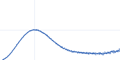
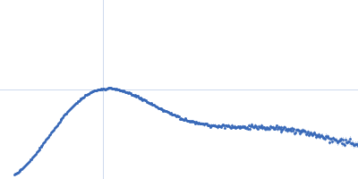
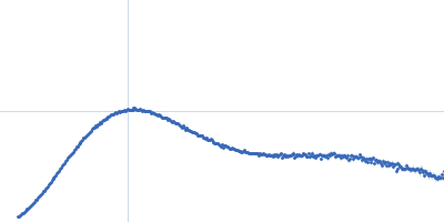
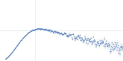
| Sample: |
Truncated P5abc subdomain from tetrahymena ribozyme monomer, 18 kDa RNA
|
| Buffer: |
20mM KCl 0.25mM MgCl2 10mM KMOPS 20uM EDTA, pH: 7 |
| Experiment: |
SAXS
data collected at G1, Cornell High Energy Synchrotron Source (CHESS) on 2016 Jun 17
|
Revealing the distinct folding phases of an RNA three-helix junction.
Nucleic Acids Res 46(14):7354-7365 (2018)
Plumridge A, Katz AM, Calvey GD, Elber R, Kirmizialtin S, Pollack L
|
| RgGuinier |
2.4 |
nm |
| Dmax |
7.6 |
nm |
|
|
|
|
|
|
|
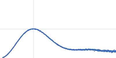
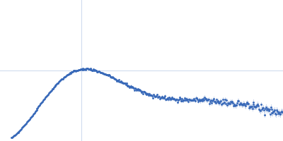
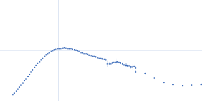
| Sample: |
Truncated P5abc subdomain from tetrahymena ribozyme monomer, 18 kDa RNA
|
| Buffer: |
0.5mM MgCl2 20mM KCl 10mM KMOPS 20uM EDTA, pH: 7 |
| Experiment: |
SAXS
data collected at G1, Cornell High Energy Synchrotron Source (CHESS) on 2016 Jun 17
|
Revealing the distinct folding phases of an RNA three-helix junction.
Nucleic Acids Res 46(14):7354-7365 (2018)
Plumridge A, Katz AM, Calvey GD, Elber R, Kirmizialtin S, Pollack L
|
| RgGuinier |
2.3 |
nm |
| Dmax |
7.2 |
nm |
|
|
|
|
|
|
|
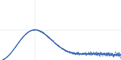
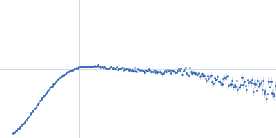
| Sample: |
Truncated P5abc subdomain from tetrahymena ribozyme monomer, 18 kDa RNA
|
| Buffer: |
1mM MgCl2 20mM KCl 10mM KMOPS 20uM EDTA, pH: 7 |
| Experiment: |
SAXS
data collected at G1, Cornell High Energy Synchrotron Source (CHESS) on 2016 Jun 17
|
Revealing the distinct folding phases of an RNA three-helix junction.
Nucleic Acids Res 46(14):7354-7365 (2018)
Plumridge A, Katz AM, Calvey GD, Elber R, Kirmizialtin S, Pollack L
|
| RgGuinier |
2.2 |
nm |
| Dmax |
7.2 |
nm |
|
|
|
|
|
|
|
| Sample: |
Lipid A export ATP-binding/permease protein MsbA dimer, 133 kDa Escherichia coli protein
Membrane scaffold protein 1D1 (deuterated, 75%) dimer, 49 kDa protein
1-palmitoyl-2-palmitoleoyl-sn-glycero-3-phosphocholine (deuteration: 78% head, 92% acyl), 1 kDa Escherichia coli
|
| Buffer: |
30 mM Tris, 150 mM NaCl, 1 mM ADP, pH: 7.5 |
| Experiment: |
SANS
data collected at D11, ILL on 2017 Mar 9
|
Conformational States of ABC Transporter MsbA in a Lipid Environment Investigated by Small-Angle Scattering Using Stealth Carrier Nanodiscs.
Structure 26(8):1072-1079.e4 (2018)
Josts I, Nitsche J, Maric S, Mertens HD, Moulin M, Haertlein M, Prevost S, Svergun DI, Busch S, Forsyth VT, Tidow H
|
| RgGuinier |
3.9 |
nm |
| Dmax |
12.5 |
nm |
| VolumePorod |
173 |
nm3 |
|
|
|
|
|
|
|
| Sample: |
Lipid A export ATP-binding/permease protein MsbA dimer, 133 kDa Escherichia coli protein
Membrane scaffold protein 1D1 (deuterated, 75%) dimer, 49 kDa protein
1-palmitoyl-2-palmitoleoyl-sn-glycero-3-phosphocholine (deuteration: 78% head, 92% acyl), 1 kDa Escherichia coli
|
| Buffer: |
30 mM Tris, 150 mM NaCl, pH: 7.5 |
| Experiment: |
SAXS
data collected at EMBL P12, PETRA III on 2017 Sep 8
|
Conformational States of ABC Transporter MsbA in a Lipid Environment Investigated by Small-Angle Scattering Using Stealth Carrier Nanodiscs.
Structure 26(8):1072-1079.e4 (2018)
Josts I, Nitsche J, Maric S, Mertens HD, Moulin M, Haertlein M, Prevost S, Svergun DI, Busch S, Forsyth VT, Tidow H
|
| RgGuinier |
4.8 |
nm |
| Dmax |
16.0 |
nm |
| VolumePorod |
607 |
nm3 |
|
|
|
|
|
|
|
| Sample: |
Nucleotide Binding Domain of Lipid A export ATP-binding/permease protein MsbA monomer, 27 kDa Escherichia coli protein
|
| Buffer: |
30 mM Tris, 150 mM NaCl, 0.5 mM TCEP, pH: 7.5 |
| Experiment: |
SAXS
data collected at EMBL P12, PETRA III on 2017 May 30
|
Conformational States of ABC Transporter MsbA in a Lipid Environment Investigated by Small-Angle Scattering Using Stealth Carrier Nanodiscs.
Structure 26(8):1072-1079.e4 (2018)
Josts I, Nitsche J, Maric S, Mertens HD, Moulin M, Haertlein M, Prevost S, Svergun DI, Busch S, Forsyth VT, Tidow H
|
| RgGuinier |
2.2 |
nm |
| Dmax |
7.3 |
nm |
| VolumePorod |
47 |
nm3 |
|
|
|
|
|
|
|
| Sample: |
Nucleotide Binding Domain of Lipid A export ATP-binding/permease protein MsbA monomer, 27 kDa Escherichia coli protein
|
| Buffer: |
30 mM Tris, 150 mM NaCl, 0.5 mM TCEP, 1 mM ADP, pH: 7.5 |
| Experiment: |
SAXS
data collected at EMBL P12, PETRA III on 2017 May 30
|
Conformational States of ABC Transporter MsbA in a Lipid Environment Investigated by Small-Angle Scattering Using Stealth Carrier Nanodiscs.
Structure 26(8):1072-1079.e4 (2018)
Josts I, Nitsche J, Maric S, Mertens HD, Moulin M, Haertlein M, Prevost S, Svergun DI, Busch S, Forsyth VT, Tidow H
|
| RgGuinier |
2.1 |
nm |
| Dmax |
7.3 |
nm |
| VolumePorod |
50 |
nm3 |
|
|
|
|
|
|
|
| Sample: |
Ferredoxin Protease monomer, 101 kDa Pectobacterium atrosepticum SCRI1043 protein
|
| Buffer: |
20 mM Tris, 150 mM NaCl, 0.03 % NaN3, 5.0 % glycerol, pH: 7.8 |
| Experiment: |
SAXS
data collected at SAXS/WAXS, Australian Synchrotron on 2017 Apr 6
|
FusC, a member of the M16 protease family acquired by bacteria for iron piracy against plants.
PLoS Biol 16(8):e2006026 (2018)
Grinter R, Hay ID, Song J, Wang J, Teng D, Dhanesakaran V, Wilksch JJ, Davies MR, Littler D, Beckham SA, Henderson IR, Strugnell RA, Dougan G, Lithgow T
|
| RgGuinier |
3.7 |
nm |
| Dmax |
12.8 |
nm |
| VolumePorod |
152 |
nm3 |
|
|
|
|
|
|
|
| Sample: |
Ferredoxin protease E83A mutant monomer, 101 kDa Pectobacterium atrosepticum SCRI1043 protein
|
| Buffer: |
20 mM Tris HCl, 150 nM NaCl, 0.02 % NaN3, 5% glycerol, pH: 7.8 |
| Experiment: |
SAXS
data collected at SAXS/WAXS, Australian Synchrotron on 2017 Apr 6
|
FusC, a member of the M16 protease family acquired by bacteria for iron piracy against plants.
PLoS Biol 16(8):e2006026 (2018)
Grinter R, Hay ID, Song J, Wang J, Teng D, Dhanesakaran V, Wilksch JJ, Davies MR, Littler D, Beckham SA, Henderson IR, Strugnell RA, Dougan G, Lithgow T
|
| RgGuinier |
3.7 |
nm |
| Dmax |
12.7 |
nm |
| VolumePorod |
152 |
nm3 |
|
|
|
|
|
|
|
| Sample: |
Ferredoxin Protease monomer, 101 kDa Pectobacterium atrosepticum SCRI1043 protein
Arabidopsis ferredoxin 2 monomer, 11 kDa Arabidopsis thaliana protein
|
| Buffer: |
20 mM Tris HCl, 150 nM NaCl, 0.02 % NaN3, 5% glycerol, pH: 7.8 |
| Experiment: |
SAXS
data collected at SAXS/WAXS, Australian Synchrotron on 2017 Apr 6
|
FusC, a member of the M16 protease family acquired by bacteria for iron piracy against plants.
PLoS Biol 16(8):e2006026 (2018)
Grinter R, Hay ID, Song J, Wang J, Teng D, Dhanesakaran V, Wilksch JJ, Davies MR, Littler D, Beckham SA, Henderson IR, Strugnell RA, Dougan G, Lithgow T
|
| RgGuinier |
3.7 |
nm |
| Dmax |
13.2 |
nm |
| VolumePorod |
156 |
nm3 |
|
|


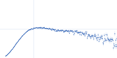

1-palmitoyl-2-palmitoleoyl-sn-glycero-3-phosphocholine (deuteration: 78% head, 92% acyl) experimental SAS data")
1-palmitoyl-2-palmitoleoyl-sn-glycero-3-phosphocholine (deuteration: 78% head, 92% acyl) experimental SAS data")